


 癌症的基本特征包括细胞增殖、血管生成、迁移、凋亡逃避机制和细胞永生等。找到癌症发生过程中这些通路的关键标记物和对应的抗体用于检测至关重要。
 为您推荐一个泛素化位点预测神器——泛素化分析工具,可以为您的蛋白的泛素化位点作出预测和评分。
 细胞自噬受体图形绘图工具为你的蛋白的细胞受体结合位点作出预测和评分,识别结合到自噬通路中的蛋白是非常重要的,便于让我们理解自噬在正常生理、病理过程中的作用,如发育、细胞分化、神经退化性疾病、压力条件下、感染和癌症。
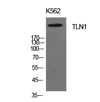
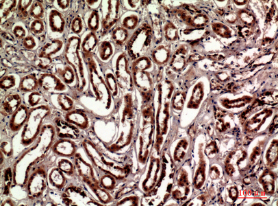
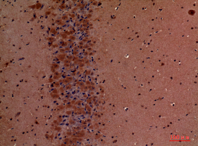
Talin-1 Polyclonal Antibody
- 产品详情
- 实验流程
- 背景知识
Application
| WB, IHC-P, IF, ICC, E |
|---|---|
| Primary Accession | Q9Y490 |
| Reactivity | Human, Mouse, Rat |
| Host | Rabbit |
| Clonality | Polyclonal |
| Calculated MW | 269767 Da |
| Gene ID | 7094 |
|---|---|
| Other Names | TLN1; KIAA1027; TLN; Talin-1 |
| Dilution | WB~~Western Blot: 1/500 - 1/2000. IHC-p: 1:100-1:300. ELISA: 1/10000. Not yet tested in other applications. IHC-P~~Western Blot: 1/500 - 1/2000. IHC-p: 1:100-1:300. ELISA: 1/10000. Not yet tested in other applications. IF~~1:50~200 ICC~~N/A E~~N/A |
| Format | Liquid in PBS containing 50% glycerol, 0.5% BSA and 0.09% (W/V) sodium azide. |
| Storage Conditions | -20℃ |
| Name | TLN1 |
|---|---|
| Synonyms | KIAA1027, TLN |
| Function | High molecular weight cytoskeletal protein concentrated at regions of cell-matrix and cell-cell contacts. Involved in connections of major cytoskeletal structures to the plasma membrane. With KANK1 co- organize the assembly of cortical microtubule stabilizing complexes (CMSCs) positioned to control microtubule-actin crosstalk at focal adhesions (FAs) rims. |
| Cellular Location | Cell projection, ruffle membrane; Peripheral membrane protein; Cytoplasmic side. Cytoplasm, cytoskeleton. Cell surface {ECO:0000250|UniProtKB:P26039}. Cell junction, focal adhesion {ECO:0000250|UniProtKB:P26039}. Note=Colocalizes with LAYN at the membrane ruffles. Localized preferentially in focal adhesions than fibrillar adhesions (By similarity). |
| Tissue Location | [Isoform 2]: Expressed at low to non-detectable levels in many tissues but highly expressed in skin and pancreas with other tissues including kidney cortex, endocervix, testis, pituitary, liver, and spleen also showing robust expression |
Research Areas
For Research Use Only. Not For Use In Diagnostic Procedures.
Application Protocols
Provided below are standard protocols that you may find useful for product applications.
BACKGROUND
Probably involved in connections of major cytoskeletal structures to the plasma membrane. High molecular weight cytoskeletal protein concentrated at regions of cell-substratum contact and, in lymphocytes, at cell-cell contacts (By similarity).
终于等到您。ABCEPTA(百远生物)抗体产品。
点击下方“我要评价 ”按钮提交您的反馈信息,您的反馈和评价是我们最宝贵的财富之一,
我们将在1-3个工作日内处理您的反馈信息。
如有疑问,联系:0512-88856768 tech-china@abcepta.com.
¥ 1,500.00
Cat# AP73759


