


 癌症的基本特征包括细胞增殖、血管生成、迁移、凋亡逃避机制和细胞永生等。找到癌症发生过程中这些通路的关键标记物和对应的抗体用于检测至关重要。
 为您推荐一个泛素化位点预测神器——泛素化分析工具,可以为您的蛋白的泛素化位点作出预测和评分。
 细胞自噬受体图形绘图工具为你的蛋白的细胞受体结合位点作出预测和评分,识别结合到自噬通路中的蛋白是非常重要的,便于让我们理解自噬在正常生理、病理过程中的作用,如发育、细胞分化、神经退化性疾病、压力条件下、感染和癌症。
CLDN7 Recombinant Rabbit mAb
CLDN7 Recombinant Rabbit mAb
- 产品详情
- 实验流程
- 背景知识
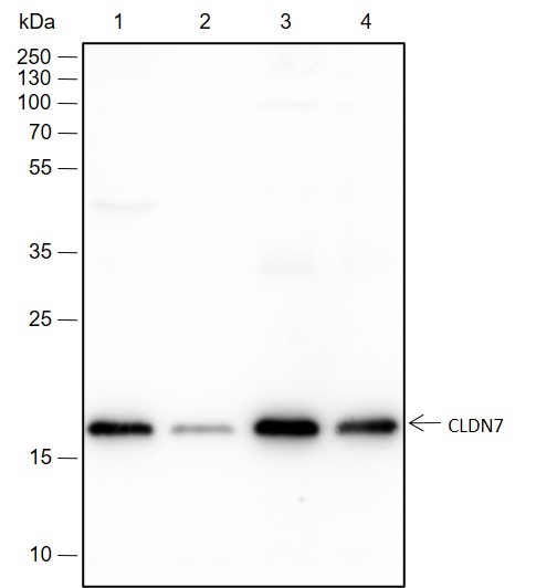
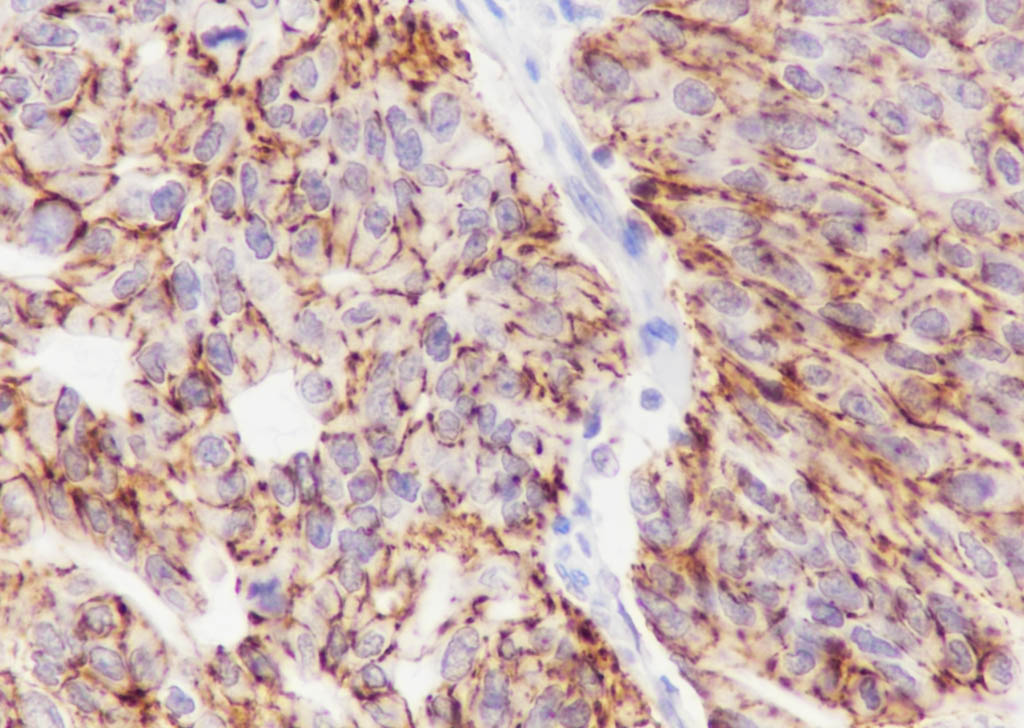
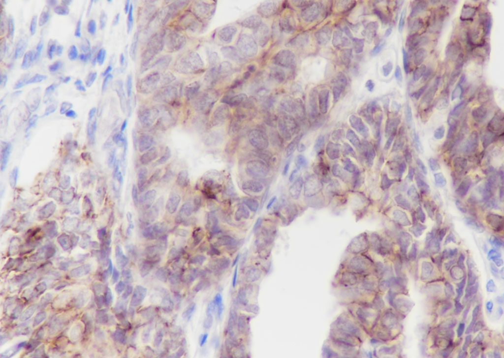
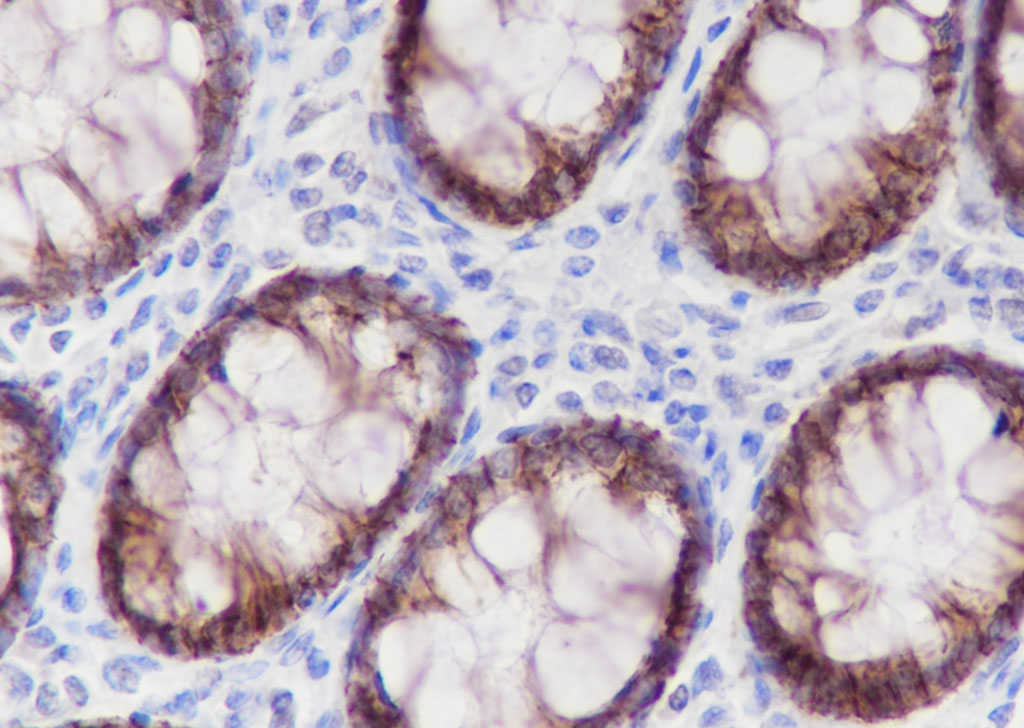
Application
| WB, IHC-P, IHC-F, IF |
|---|---|
| Host | Rabbit |
| Clonality | Recombinant |
| Calculated MW | 22 KDa |
| Physical State | Liquid |
| Isotype | IgG |
| Purity | affinity purified by Protein A |
| Buffer | 0.01M TBS (pH7.4) with 1% BSA, 0.02% Proclin300 and 50% Glycerol. |
| SUBCELLULAR LOCATION | Cell membrane. Lateral cell membrane. Cell junction > tight junction. Co-localizes with EPCAM at the lateral cell membrane and tight junction. |
| SIMILARITY | Belongs to the claudin family. |
| SUBUNIT | Directly interacts with TJP1/ZO-1, TJP2/ZO-2 and TJP3/ZO-3 (By similarity). The phosphorylated form interacts with EPCAM. |
| Post-translational modifications | Phosphorylated. |
| Important Note | This product as supplied is intended for research use only, not for use in human, therapeutic or diagnostic applications. |
| Background Descriptions | This gene encodes a member of the claudin family. Claudins are integral membrane proteins and components of tight junction strands. Tight junction strands serve as a physical barrier to prevent solutes and water from passing freely through the paracellular space between epithelial or endothelial cell sheets, and also play critical roles in maintaining cell polarity and signal transductions. Differential expression of this gene has been observed in different types of malignancies, including breast cancer, ovarian cancer, hepatocellular carcinomas, urinary tumors, prostate cancer, lung cancer, head and neck cancers, thyroid carcinomas, etc.. Alternatively spliced transcript variants encoding different isoforms have been found.[provided by RefSeq, May 2010] |
| Target/Specificity | Expressed in kidney, lung and prostate. Isoform 1 seems to be predominant, except in some normal prostate samples, where isoform 2 is the major form. Down-regulated in breast cancers, including ductal carcinoma in situ (DCIS), lobular carcinoma in situ (LCIS) and invasive ductal carcinoma (IDC) (at protein level), as well as in several cancer cell lines. Loss of expression correlates with histological grade, occurring predominantly in high-grade lesions. |
|---|---|
| Dilution | WB=1:200-1000,IHC-P=1:20-100,IHC-F=1:20-100,IF=1:20-100 |
| Format | 0.01M TBS(pH7.4) with 1% BSA, 0.09% (W/V) sodium azide and 50% Glyce |
| Storage | Store at -20 °C for one year. Avoid repeated freeze/thaw cycles. When reconstituted in sterile pH 7.4 0.01M PBS or diluent of antibody the antibody is stable for at least two weeks at 2-4 °C. |
Research Areas
For Research Use Only. Not For Use In Diagnostic Procedures.
Application Protocols
Provided below are standard protocols that you may find useful for product applications.
BACKGROUND
This product as supplied is intended for research use only, not for use in human, therapeutic or diagnostic applications.
终于等到您。ABCEPTA(百远生物)抗体产品。
点击下方“我要评价 ”按钮提交您的反馈信息,您的反馈和评价是我们最宝贵的财富之一,
我们将在1-3个工作日内处理您的反馈信息。
如有疑问,联系:0512-88856768 tech-china@abcepta.com.


