


 癌症的基本特征包括细胞增殖、血管生成、迁移、凋亡逃避机制和细胞永生等。找到癌症发生过程中这些通路的关键标记物和对应的抗体用于检测至关重要。
 为您推荐一个泛素化位点预测神器——泛素化分析工具,可以为您的蛋白的泛素化位点作出预测和评分。
 细胞自噬受体图形绘图工具为你的蛋白的细胞受体结合位点作出预测和评分,识别结合到自噬通路中的蛋白是非常重要的,便于让我们理解自噬在正常生理、病理过程中的作用,如发育、细胞分化、神经退化性疾病、压力条件下、感染和癌症。
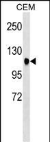
ATXN7 Antibody (Center)
Affinity Purified Rabbit Polyclonal Antibody (Pab)
- 产品详情
- 实验流程
- 背景知识
Application
| WB, E |
|---|---|
| Primary Accession | O15265 |
| Other Accession | Q8R4I1, NP_000324.1, NP_001170858.1 |
| Reactivity | Human |
| Predicted | Mouse |
| Host | Rabbit |
| Clonality | Polyclonal |
| Isotype | Rabbit IgG |
| Calculated MW | 95451 Da |
| Antigen Region | 354-381 aa |
| Gene ID | 6314 |
|---|---|
| Other Names | Ataxin-7, Spinocerebellar ataxia type 7 protein, ATXN7, SCA7 |
| Target/Specificity | This ATXN7 antibody is generated from rabbits immunized with a KLH conjugated synthetic peptide between 354-381 amino acids from the Central region of human ATXN7. |
| Dilution | WB~~1:1000 E~~Use at an assay dependent concentration. |
| Format | Purified polyclonal antibody supplied in PBS with 0.09% (W/V) sodium azide. This antibody is purified through a protein A column, followed by peptide affinity purification. |
| Storage | Maintain refrigerated at 2-8°C for up to 2 weeks. For long term storage store at -20°C in small aliquots to prevent freeze-thaw cycles. |
| Precautions | ATXN7 Antibody (Center) is for research use only and not for use in diagnostic or therapeutic procedures. |
| Name | ATXN7 |
|---|---|
| Synonyms | SCA7 {ECO:0000303|PubMed:9288099} |
| Function | Acts as a component of the SAGA (aka STAGA) transcription coactivator-HAT complex (PubMed:15932940, PubMed:18206972). Mediates the interaction of SAGA complex with the CRX and is involved in CRX- dependent gene activation (PubMed:15932940, PubMed:18206972). Probably involved in tethering the deubiquitination module within the SAGA complex (PubMed:24493646). Necessary for microtubule cytoskeleton stabilization (PubMed:22100762). Involved in neurodegeneration (PubMed:9288099). |
| Cellular Location | [Isoform a]: Nucleus. Nucleus, nucleolus. Nucleus matrix. Cytoplasm, cytoskeleton. Note=In addition to a diffuse distribution throughout the nucleus, it is associated with the nuclear matrix and the nucleolus (PubMed:10441328). It is able to shuttle between the nucleus and cytoplasm (PubMed:16314424) |
| Tissue Location | [Isoform a]: Isoform a is expressed in CNS, but is expressed predominantly in the peripherical tissues |
For Research Use Only. Not For Use In Diagnostic Procedures.
Provided below are standard protocols that you may find useful for product applications.
BACKGROUND
The autosomal dominant cerebellar ataxias (ADCA) are a heterogeneous group of neurodegenerative disorders characterized by progressive degeneration of the cerebellum, brain stem and spinal cord. Clinically, ADCA has been divided into three groups: ADCA types I-III. ADCAI is genetically heterogeneous, with five genetic loci, designated spinocerebellar ataxia (SCA) 1, 2, 3, 4 and 6, being assigned to five different chromosomes. ADCAII, which always presents with retinal degeneration (SCA7), and ADCAIII often referred to as the 'pure' cerebellar syndrome (SCA5), are most likely homogeneous disorders. Several SCA genes have been cloned and shown to contain CAG repeats in their coding regions. ADCA is caused by the expansion of the CAG repeats, producing an elongated polyglutamine tract in the corresponding protein. The expanded repeats are variable in size and unstable, usually increasing in size when transmitted to successive generations. This locus has been mapped to chromosome 3, and it has been determined that the diseased allele associated with spinocerebellar ataxia-7 contains 38-130 CAG repeats (near the N-terminus), compared to 7-17 in the normal allele. The encoded protein is a component of the SPT3/TAF9/GCN5 acetyltransferase (STAGA) and TBP-free TAF-containing (TFTC) chromatin remodeling complexes, and it thus plays a role in transcriptional regulation. Alternative splicing results in multiple transcript variants.
REFERENCES
Bonnet, J., et al. EMBO Rep. 11(8):612-618(2010)
Han, Y., et al. Neurol India 58(4):622-626(2010)
Chou, A.H., et al. Neurochem. Int. 56(2):329-339(2010)
Mookerjee, S., et al. J. Neurosci. 29(48):15134-15144(2009)
Freund, A.A., et al. Arq Neuropsiquiatr 67(4):1124-1132(2009)
终于等到您。ABCEPTA(百远生物)抗体产品。
点击下方“我要评价 ”按钮提交您的反馈信息,您的反馈和评价是我们最宝贵的财富之一,
我们将在1-3个工作日内处理您的反馈信息。
如有疑问,联系:0512-88856768 tech-china@abcepta.com.


