


 癌症的基本特征包括细胞增殖、血管生成、迁移、凋亡逃避机制和细胞永生等。找到癌症发生过程中这些通路的关键标记物和对应的抗体用于检测至关重要。
 为您推荐一个泛素化位点预测神器——泛素化分析工具,可以为您的蛋白的泛素化位点作出预测和评分。
 细胞自噬受体图形绘图工具为你的蛋白的细胞受体结合位点作出预测和评分,识别结合到自噬通路中的蛋白是非常重要的,便于让我们理解自噬在正常生理、病理过程中的作用,如发育、细胞分化、神经退化性疾病、压力条件下、感染和癌症。
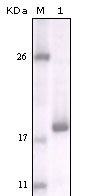
DDR2 Antibody
Purified Mouse Monoclonal Antibody
- 产品详情
- 实验流程
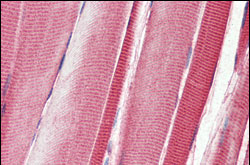
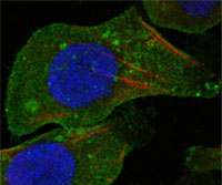
Application
| WB, IHC, ICC, E |
|---|---|
| Primary Accession | Q16832 |
| Reactivity | Human |
| Host | Mouse |
| Clonality | Monoclonal |
| Clone Names | 3B11E4 |
| Isotype | IgG2a |
| Calculated MW | 96736 Da |
| Description | DDR2 (discoidin domain receptor family, member 2) is one of the largest families of proteins in eukaryotes. The family has been classified in 8 major groups based on sequence comparison of their tyrosine (PTK) or serine/ threonine (STK) kinase catalytic domains. Receptor tyrosine kinases (RTKs) play a key role in the communication of cells with their microenvironment. These molecules are involved in the regulation of cell growth, differentiation, and metabolism. In several cases the biochemical mechanism by which RTKs transduce signals across the membrane has been shown to be ligand induced receptor oligomerization and subsequent intracellular phosphorylation. This autophosphorylation leads to phosphorylation of cytosolic targets as well as association with other molecules, which are involved in pleiotropic effects of signal transduction. RTKs have a tripartite structure with extracellular, transmembrane, and cytoplasmic regions. This gene encodes a member of a novel subclass of RTKs and contains a distinct extracellular region encompassing a factor VIII-like domain. Alternative splicing in the 5' UTR results in multiple transcript variants encoding the same protein. |
| Immunogen | Purified recombinant fragment of human DDR2 expressed in E. Coli. |
| Formulation | Ascitic fluid containing 0.03% sodium azide. |
| Gene ID | 4921 |
|---|---|
| Other Names | Discoidin domain-containing receptor 2, Discoidin domain receptor 2, 2.7.10.1, CD167 antigen-like family member B, Discoidin domain-containing receptor tyrosine kinase 2, Neurotrophic tyrosine kinase, receptor-related 3, Receptor protein-tyrosine kinase TKT, Tyrosine-protein kinase TYRO10, CD167b, DDR2, NTRKR3, TKT, TYRO10 |
| Dilution | WB~~1/500 - 1/2000 IHC~~1/200 - 1/1000 ICC~~N/A E~~N/A |
| Storage | Maintain refrigerated at 2-8°C for up to 6 months. For long term storage store at -20°C in small aliquots to prevent freeze-thaw cycles. |
| Precautions | DDR2 Antibody is for research use only and not for use in diagnostic or therapeutic procedures. |
| Name | DDR2 |
|---|---|
| Synonyms | NTRKR3, TKT, TYRO10 |
| Function | Tyrosine kinase involved in the regulation of tissues remodeling (PubMed:30449416). It functions as a cell surface receptor for fibrillar collagen and regulates cell differentiation, remodeling of the extracellular matrix, cell migration and cell proliferation. Required for normal bone development. Regulates osteoblast differentiation and chondrocyte maturation via a signaling pathway that involves MAP kinases and leads to the activation of the transcription factor RUNX2. Regulates remodeling of the extracellular matrix by up- regulation of the collagenases MMP1, MMP2 and MMP13, and thereby facilitates cell migration and tumor cell invasion. Promotes fibroblast migration and proliferation, and thereby contributes to cutaneous wound healing. |
| Cellular Location | Cell membrane; Single-pass type I membrane protein |
| Tissue Location | Detected in osteocytes, osteoblastic cells in subchondral bone, bone lining cells, tibia and cartilage (at protein level). Detected at high levels in heart and lung, and at low levels in brain, placenta, liver, skeletal muscle, pancreas, and kidney |
Research Areas
For Research Use Only. Not For Use In Diagnostic Procedures.
Application Protocols
Provided below are standard protocols that you may find useful for product applications.
REFERENCES
1. Leitinger B. Kwan AP. Matrix Biol. 2006, Aug, 25(6):355-64. Epub 2006 May 26. 2. Shyu KG. Chao YM. Wang BW. et al. Hypertension. 2005, Sep, 46(3):614-21. Epub 2005 Aug 8. 3. Neale JC. Kenny TP. Gershwin ME. Clin Dev Immunol. 2004, Jun, 11(2):157-63.
终于等到您。ABCEPTA(百远生物)抗体产品。
点击下方“我要评价 ”按钮提交您的反馈信息,您的反馈和评价是我们最宝贵的财富之一,
我们将在1-3个工作日内处理您的反馈信息。
如有疑问,联系:0512-88856768 tech-china@abcepta.com.
¥ 1,500.00
Cat# AO1124a


