


 癌症的基本特征包括细胞增殖、血管生成、迁移、凋亡逃避机制和细胞永生等。找到癌症发生过程中这些通路的关键标记物和对应的抗体用于检测至关重要。
 为您推荐一个泛素化位点预测神器——泛素化分析工具,可以为您的蛋白的泛素化位点作出预测和评分。
 细胞自噬受体图形绘图工具为你的蛋白的细胞受体结合位点作出预测和评分,识别结合到自噬通路中的蛋白是非常重要的,便于让我们理解自噬在正常生理、病理过程中的作用,如发育、细胞分化、神经退化性疾病、压力条件下、感染和癌症。
Livin Antibody
- 产品详情
- 实验流程
- 背景知识
Application
| WB, IF, E, IHC-P |
|---|---|
| Primary Accession | Q96CA5 |
| Other Accession | NP_071444, 11545910 |
| Reactivity | Human |
| Host | Rabbit |
| Clonality | Polyclonal |
| Isotype | IgG |
| Calculated MW | 32798 Da |
| Conjugate | Unconjugated |
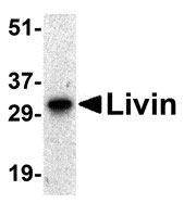
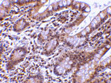
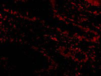
| Application Notes | Livin antibody can be used for detection of Livin by Western blot at 0.5 µg/mL. A band at 33 kDa can be detected. Antibody can also be used for immunohistochemistry starting at 5 µg/mL. For immunofluorescence start at 20 µg/mL. |
| Gene ID | 79444 |
|---|---|
| Other Names | Livin Antibody: KIAP, LIVIN, MLIAP, RNF50, ML-IAP, KIAP, UNQ5800/PRO19607/PRO21344, Baculoviral IAP repeat-containing protein 7, Kidney inhibitor of apoptosis protein, baculoviral IAP repeat-containing 7 |
| Target/Specificity | BIRC7; A lower but much weaker band at 30 kDa was detected in Raji cell lysate, which may represent the short form of Livin. |
| Reconstitution & Storage | Livin antibody can be stored at 4℃ for three months and -20℃, stable for up to one year. As with all antibodies care should be taken to avoid repeated freeze thaw cycles. Antibodies should not be exposed to prolonged high temperatures. |
| Precautions | Livin Antibody is for research use only and not for use in diagnostic or therapeutic procedures. |
| Name | BIRC7 |
|---|---|
| Synonyms | KIAP, LIVIN, MLIAP, RNF50 |
| Function | Apoptotic regulator capable of exerting proapoptotic and anti-apoptotic activities and plays crucial roles in apoptosis, cell proliferation, and cell cycle control (PubMed:11024045, PubMed:11084335, PubMed:11162435, PubMed:16729033, PubMed:17294084). Its anti-apoptotic activity is mediated through the inhibition of CASP3, CASP7 and CASP9, as well as by its E3 ubiquitin-protein ligase activity (PubMed:11024045, PubMed:16729033). As it is a weak caspase inhibitor, its anti-apoptotic activity is thought to be due to its ability to ubiquitinate DIABLO/SMAC targeting it for degradation thereby promoting cell survival (PubMed:16729033). May contribute to caspase inhibition, by blocking the ability of DIABLO/SMAC to disrupt XIAP/BIRC4-caspase interactions (PubMed:16729033). Protects against apoptosis induced by TNF or by chemical agents such as adriamycin, etoposide or staurosporine (PubMed:11084335, PubMed:11162435, PubMed:11865055). Suppression of apoptosis is mediated by activation of MAPK8/JNK1, and possibly also of MAPK9/JNK2 (PubMed:11865055). This activation depends on TAB1 and MAP3K7/TAK1 (PubMed:11865055). In vitro, inhibits CASP3 and proteolytic activation of pro-CASP9 (PubMed:11024045). |
| Cellular Location | Nucleus. Cytoplasm. Golgi apparatus. Note=Nuclear, and in a filamentous pattern throughout the cytoplasm. Full-length livin is detected exclusively in the cytoplasm, whereas the truncated form (tLivin) is found in the peri-nuclear region with marked localization to the Golgi apparatus; the accumulation of tLivin in the nucleus shows positive correlation with the increase in apoptosis |
| Tissue Location | Isoform 1 and isoform 2 are expressed at very low levels or not detectable in most adult tissues. Detected in adult heart, placenta, lung, lymph node, spleen and ovary, and in several carcinoma cell lines. Isoform 2 is detected in fetal kidney, heart and spleen, and at lower levels in adult brain, skeletal muscle and peripheral blood leukocytes |
For Research Use Only. Not For Use In Diagnostic Procedures.
Provided below are standard protocols that you may find useful for product applications.
BACKGROUND
Livin Antibody: Apoptosis, or programmed cell death, is related to many diseases, such as cancer. Apoptosis is triggered by a variety of stimuli including members in the TNF family and prevented by the inhibitor of apoptosis (IAP) proteins. IAP proteins form a conserved gene family that binds to and inhibits cell death proteases. A novel member in the IAP protein family was recently identified and designated Livin and KIAP for kidney IAP. Livin/XIAP contains a single baculoviral IAP repeat (BIR) domain and a RING finger domain and has two isoforms termed Livin-alpha and Livin-beta. Transfection of Livin in cells resulted in protection from apoptosis induced by FADD, BAX, RIP, RIP3 and DR6. Livin has direct interaction with several caspases including caspase-3, -7, and -9. Livin inhibits the activation of caspase-9 induced by Apaf-1, cytochrome c, and dATP. The two isoforms of Livin appear to have different functions and tissue distributions.
REFERENCES
Kasof GM, Gomes BC. Livin, a novel inhibitor of apoptosis protein family member. J Biol Chem. 2001;276(5):3238-46.
Lin JH, Deng G, Huang Q, Morser J. KIAP, a novel member of the inhibitor of apoptosis protein family. Biochem Biophys Res Commun. 2000;279(3):820-31.
Ashhab Y, Alian A, Polliack A, Panet A, Yehuda DB. Two splicing variants of a new inhibitor of apoptosis gene with different biological properties and tissue distribution pattern. FEBS Lett. 2001;495(1-2):56-60.
终于等到您。ABCEPTA(百远生物)抗体产品。
点击下方“我要评价 ”按钮提交您的反馈信息,您的反馈和评价是我们最宝贵的财富之一,
我们将在1-3个工作日内处理您的反馈信息。
如有疑问,联系:0512-88856768 tech-china@abcepta.com.


