


 癌症的基本特征包括细胞增殖、血管生成、迁移、凋亡逃避机制和细胞永生等。找到癌症发生过程中这些通路的关键标记物和对应的抗体用于检测至关重要。
 为您推荐一个泛素化位点预测神器——泛素化分析工具,可以为您的蛋白的泛素化位点作出预测和评分。
 细胞自噬受体图形绘图工具为你的蛋白的细胞受体结合位点作出预测和评分,识别结合到自噬通路中的蛋白是非常重要的,便于让我们理解自噬在正常生理、病理过程中的作用,如发育、细胞分化、神经退化性疾病、压力条件下、感染和癌症。
Anti-IFNAR1 Reference Antibody (Faralimomab)
Recombinant Antibody
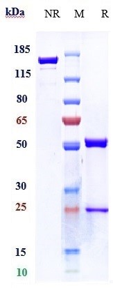
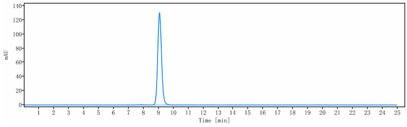
- 产品详情
- 实验流程
Application
| FC, Kinetics, Animal Model |
|---|---|
| Primary Accession | P17181 |
| Reactivity | Human |
| Clonality | Monoclonal |
| Isotype | mIgG1 |
| Calculated MW | 63525 Da |
| Target/Specificity | IFNAR1 |
|---|---|
| Endotoxin | < 0.001EU/ µg,determined by LAL method. |
| Conjugation | Unconjugated |
| Expression system | CHO Cell |
| Format | Purified monoclonal antibody supplied in PBS, pH6.0, without preservative.This antibody is purified through a protein A column. |
| Name | IFNAR1 |
|---|---|
| Synonyms | IFNAR |
| Function | Together with IFNAR2, forms the heterodimeric receptor for type I interferons (including interferons alpha, beta, epsilon, omega and kappa) (PubMed:10049744, PubMed:14532120, PubMed:15337770, PubMed:2153461, PubMed:21854986, PubMed:24075985, PubMed:31270247, PubMed:33252644, PubMed:35442418, PubMed:7813427). Type I interferon binding activates the JAK-STAT signaling cascade, resulting in transcriptional activation or repression of interferon-regulated genes that encode the effectors of the interferon response (PubMed:10049744, PubMed:21854986, PubMed:7665574). Mechanistically, type I interferon- binding brings the IFNAR1 and IFNAR2 subunits into close proximity with one another, driving their associated Janus kinases (JAKs) (TYK2 bound to IFNAR1 and JAK1 bound to IFNAR2) to cross-phosphorylate one another (PubMed:21854986, PubMed:32972995, PubMed:7665574, PubMed:7813427). The activated kinases phosphorylate specific tyrosine residues on the intracellular domains of IFNAR1 and IFNAR2, forming docking sites for the STAT transcription factors (PubMed:21854986, PubMed:32972995, PubMed:7526154, PubMed:7665574, PubMed:7813427). STAT proteins are then phosphorylated by the JAKs, promoting their translocation into the nucleus to regulate expression of interferon-regulated genes (PubMed:19561067, PubMed:21854986, PubMed:32972995, PubMed:7665574, PubMed:7813427, PubMed:9121453). Can also act independently of IFNAR2: form an active IFNB1 receptor by itself and activate a signaling cascade that does not involve activation of the JAK-STAT pathway (By similarity). |
| Cellular Location | [Isoform 1]: Cell membrane; Single-pass type I membrane protein. Late endosome. Lysosome. Note=Interferon binding triggers internalization of the receptor from the cell membrane into endosomes and then into lysosomes. |
| Tissue Location | IFN receptors are present in all tissues and even on the surface of most IFN-resistant cells. Isoform 1, isoform 2 and isoform 3 are expressed in the IFN-alpha sensitive myeloma cell line U266B1. Isoform 2 and isoform 3 are expressed in the IFN-alpha resistant myeloma cell line U266R. Isoform 1 is not expressed in IFN- alpha resistant myeloma cell line U266R. |
Research Areas
For Research Use Only. Not For Use In Diagnostic Procedures.
Application Protocols
Provided below are standard protocols that you may find useful for product applications.
终于等到您。ABCEPTA(百远生物)抗体产品。
点击下方“我要评价 ”按钮提交您的反馈信息,您的反馈和评价是我们最宝贵的财富之一,
我们将在1-3个工作日内处理您的反馈信息。
如有疑问,联系:0512-88856768 tech-china@abcepta.com.
¥ 1,500.00
Cat# APR10945


