


 癌症的基本特征包括细胞增殖、血管生成、迁移、凋亡逃避机制和细胞永生等。找到癌症发生过程中这些通路的关键标记物和对应的抗体用于检测至关重要。
 为您推荐一个泛素化位点预测神器——泛素化分析工具,可以为您的蛋白的泛素化位点作出预测和评分。
 细胞自噬受体图形绘图工具为你的蛋白的细胞受体结合位点作出预测和评分,识别结合到自噬通路中的蛋白是非常重要的,便于让我们理解自噬在正常生理、病理过程中的作用,如发育、细胞分化、神经退化性疾病、压力条件下、感染和癌症。
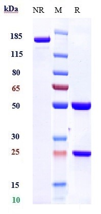
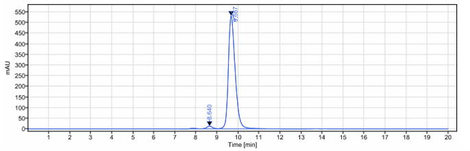
Anti-CXADR Reference Antibody (Med. Bio. Labs patent anti-CXADR)
Recombinant Antibody
- 产品详情
- 实验流程
Application
| FC, Kinetics, Animal Model |
|---|---|
| Primary Accession | P78310 |
| Reactivity | Human |
| Clonality | Monoclonal |
| Isotype | IgG2SA |
| Calculated MW | 40030 Da |
| Target/Specificity | CXADR |
|---|---|
| Endotoxin | < 0.001EU/ µg,determined by LAL method. |
| Conjugation | Unconjugated |
| Expression system | CHO Cell |
| Format | Purified monoclonal antibody supplied in PBS, pH6.0, without preservative.This antibody is purified through a protein A column. |
| Name | CXADR |
|---|---|
| Synonyms | CAR |
| Function | Component of the epithelial apical junction complex that may function as a homophilic cell adhesion molecule and is essential for tight junction integrity. Also involved in transepithelial migration of leukocytes through adhesive interactions with JAML a transmembrane protein of the plasma membrane of leukocytes. The interaction between both receptors also mediates the activation of gamma-delta T-cells, a subpopulation of T-cells residing in epithelia and involved in tissue homeostasis and repair. Upon epithelial CXADR-binding, JAML induces downstream cell signaling events in gamma-delta T-cells through PI3- kinase and MAP kinases. It results in proliferation and production of cytokines and growth factors by T-cells that in turn stimulate epithelial tissues repair. |
| Cellular Location | [Isoform 1]: Cell membrane; Single-pass type I membrane protein. Basolateral cell membrane; Single-pass type I membrane protein. Cell junction, tight junction. Cell junction, adherens junction. Note=In epithelial cells localizes to the apical junction complex composed of tight and adherens junctions (PubMed:12297051). In airway epithelial cells localized to basolateral membrane but not to apical surface (PubMed:11316797). [Isoform 4]: Secreted |
| Tissue Location | Expressed in pancreas, brain, heart, small intestine, testis, prostate and at a lower level in liver and lung Isoform 5 is ubiquitously expressed. Isoform 3 is expressed in heart, lung and pancreas. In skeletal muscle, isoform 1 is found at the neuromuscular junction and isoform 2 is found in blood vessels. In cardiac muscle, isoform 1 and isoform 2 are found at intercalated disks. In heart expressed in subendothelial layers of the vessel wall but not in the luminal endothelial surface. Expression is elevated in hearts with dilated cardiomyopathy. |
Research Areas
For Research Use Only. Not For Use In Diagnostic Procedures.
Application Protocols
Provided below are standard protocols that you may find useful for product applications.
终于等到您。ABCEPTA(百远生物)抗体产品。
点击下方“我要评价 ”按钮提交您的反馈信息,您的反馈和评价是我们最宝贵的财富之一,
我们将在1-3个工作日内处理您的反馈信息。
如有疑问,联系:0512-88856768 tech-china@abcepta.com.
¥ 1,500.00
Cat# APR10869


