


 癌症的基本特征包括细胞增殖、血管生成、迁移、凋亡逃避机制和细胞永生等。找到癌症发生过程中这些通路的关键标记物和对应的抗体用于检测至关重要。
 为您推荐一个泛素化位点预测神器——泛素化分析工具,可以为您的蛋白的泛素化位点作出预测和评分。
 细胞自噬受体图形绘图工具为你的蛋白的细胞受体结合位点作出预测和评分,识别结合到自噬通路中的蛋白是非常重要的,便于让我们理解自噬在正常生理、病理过程中的作用,如发育、细胞分化、神经退化性疾病、压力条件下、感染和癌症。
Anti-CD98 Reference Antibody (KHK2898)
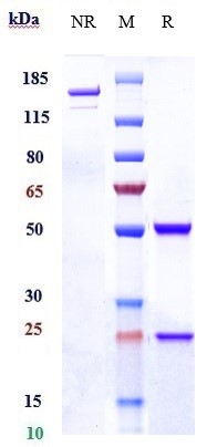
Recombinant Antibody
- 产品详情
- 实验流程
Application
| FC, Kinetics, Animal Model |
|---|---|
| Primary Accession | P08195 |
| Reactivity | Human |
| Clonality | Monoclonal |
| Isotype | IgG1 |
| Calculated MW | 67994 Da |
| Target/Specificity | CD98 |
|---|---|
| Endotoxin | < 0.001EU/ µg,determined by LAL method. |
| Conjugation | Unconjugated |
| Expression system | CHO Cell |
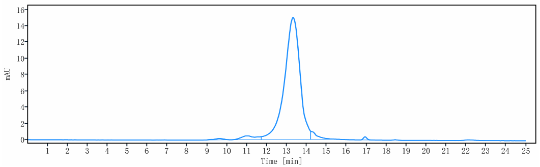
| Format | Purified monoclonal antibody supplied in PBS, pH6.0, without preservative.This antibody is purified through a protein A column. |
| Name | SLC3A2 (HGNC:11026) |
|---|---|
| Synonyms | MDU1 |
| Function | Acts as a chaperone that facilitates biogenesis and trafficking of functional transporters heterodimers to the plasma membrane. Forms heterodimer with SLC7 family transporters (SLC7A5, SLC7A6, SLC7A7, SLC7A8, SLC7A10 and SLC7A11), a group of amino-acid antiporters (PubMed:10574970, PubMed:10903140, PubMed:11557028, PubMed:30867591, PubMed:33298890, PubMed:33758168, PubMed:34880232, PubMed:9751058, PubMed:9829974, PubMed:9878049). Heterodimers function as amino acids exchangers, the specificity of the substrate depending on the SLC7A subunit. Heterodimers SLC3A2/SLC7A6 or SLC3A2/SLC7A7 mediate the uptake of dibasic amino acids (PubMed:10903140, PubMed:9829974). Heterodimer SLC3A2/SLC7A11 functions as an antiporter by mediating the exchange of extracellular anionic L-cystine and intracellular L-glutamate across the cellular plasma membrane (PubMed:34880232). SLC3A2/SLC7A10 translocates small neutral L- and D- amino acids across the plasma membrane (By similarity). SLC3A2/SLC75 or SLC3A2/SLC7A8 translocates neutral amino acids with broad specificity, thyroid hormones and L-DOPA (PubMed:10574970, PubMed:11389679, PubMed:11557028, PubMed:11564694, PubMed:11742812, PubMed:12117417, PubMed:12225859, PubMed:12716892, PubMed:15980244, PubMed:30867591, PubMed:33298890, PubMed:33758168). SLC3A2 is essential for plasma membrane localization, stability, and the transport activity of SLC7A5 and SLC7A8 (PubMed:10391915, PubMed:10574970, PubMed:11311135, PubMed:15769744, PubMed:33066406). When associated with LAPTM4B, the heterodimer SLC7A5 is recruited to lysosomes to promote leucine uptake into these organelles, and thereby mediates mTORC1 activation (PubMed:25998567). Modulates integrin-related signaling and is essential for integrin-dependent cell spreading, migration and tumor progression (PubMed:11121428, PubMed:15625115). |
| Cellular Location | Apical cell membrane. Cell membrane; Single-pass type II membrane protein. Cell junction {ECO:0000250|UniProtKB:P10852}. Lysosome membrane. Melanosome. Basolateral cell membrane {ECO:0000250|UniProtKB:P10852}. Note=Localized at the plasma membrane when associated with SLC7A5/LAT1 or SLC7A8/LAT2 (PubMed:11311135, PubMed:9751058). Localized to the apical membrane of placental syncytiotrophoblastic cells (PubMed:11742812). Recruited to lysosomes by LAPTM4B (PubMed:25998567). Identified by mass spectrometry in melanosome fractions from stage I to stage IV (PubMed:17081065) Located selectively at cell-cell adhesion sites (By similarity) Colocalized with SLC7A8/LAT2 at the basolateral membrane of kidney proximal tubules and small intestine epithelia. Expressed in both luminal and abluminal membranes of brain capillary endothelial cells (By similarity). {ECO:0000250|UniProtKB:P10852, ECO:0000269|PubMed:11311135, ECO:0000269|PubMed:11742812, ECO:0000269|PubMed:17081065, ECO:0000269|PubMed:25998567, ECO:0000269|PubMed:9751058} |
| Tissue Location | Expressed ubiquitously in all tissues tested with highest levels detected in kidney, placenta and testis and weakest level in thymus. During gestation, expression in the placenta was significantly stronger at full-term than at the mid-trimester stage Expressed in HUVECS and at low levels in resting peripheral blood T- lymphocytes and quiescent fibroblasts. Also expressed in fetal liver and in the astrocytic process of primary astrocytic gliomas. Expressed in retinal endothelial cells and in the intestinal epithelial cell line C2BBe1. |
Research Areas
For Research Use Only. Not For Use In Diagnostic Procedures.
Application Protocols
Provided below are standard protocols that you may find useful for product applications.
终于等到您。ABCEPTA(百远生物)抗体产品。
点击下方“我要评价 ”按钮提交您的反馈信息,您的反馈和评价是我们最宝贵的财富之一,
我们将在1-3个工作日内处理您的反馈信息。
如有疑问,联系:0512-88856768 tech-china@abcepta.com.
¥ 1,500.00
Cat# APR10486


