


 癌症的基本特征包括细胞增殖、血管生成、迁移、凋亡逃避机制和细胞永生等。找到癌症发生过程中这些通路的关键标记物和对应的抗体用于检测至关重要。
 为您推荐一个泛素化位点预测神器——泛素化分析工具,可以为您的蛋白的泛素化位点作出预测和评分。
 细胞自噬受体图形绘图工具为你的蛋白的细胞受体结合位点作出预测和评分,识别结合到自噬通路中的蛋白是非常重要的,便于让我们理解自噬在正常生理、病理过程中的作用,如发育、细胞分化、神经退化性疾病、压力条件下、感染和癌症。
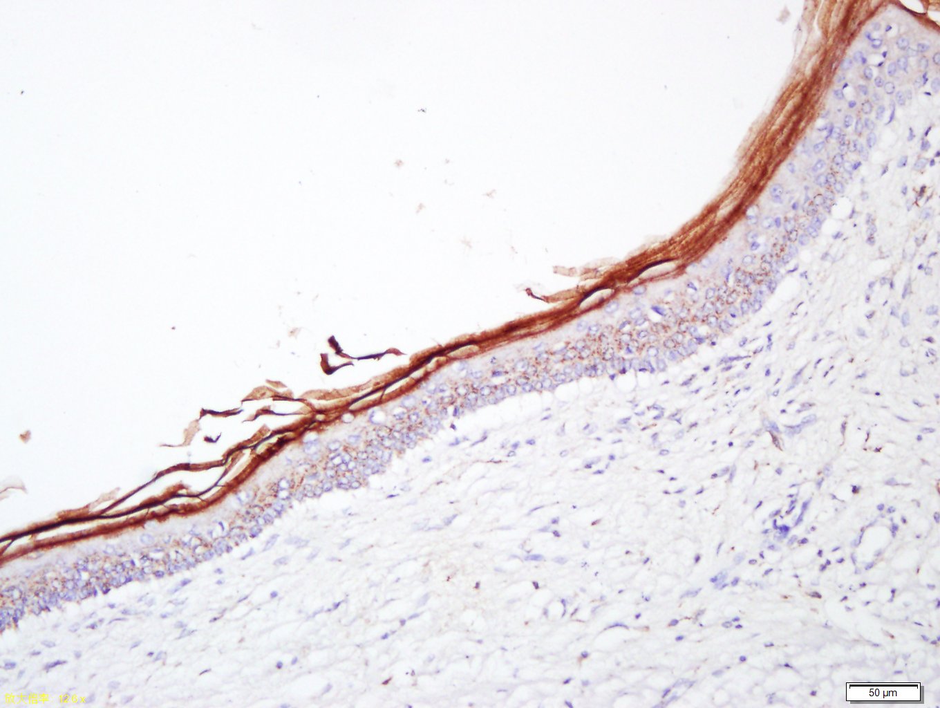
Filaggrin Rabbit pAb
Filaggrin Rabbit pAb
- 产品详情
- 实验流程
- 背景知识
Application
| IHC-P, IHC-F, IF |
|---|---|
| Primary Accession | P20930 |
| Reactivity | Human, Rat |
| Predicted | Mouse, Dog, Pig, Horse, Rabbit |
| Host | Rabbit |
| Clonality | Polyclonal |
| Calculated MW | 435170 Da |
| Physical State | Liquid |
| Immunogen | KLH conjugated synthetic peptide derived from human Filaggrin |
| Epitope Specificity | 21-150/4061 |
| Isotype | IgG |
| Purity | affinity purified by Protein A |
| Buffer | 0.01M TBS (pH7.4) with 1% BSA, 0.02% Proclin300 and 50% Glycerol. |
| SIMILARITY | Belongs to the S100-fused protein family. Contains 2 EF-hand domains. Contains 23 filaggrin repeats. |
| Post-translational modifications | Filaggrin is initially synthesized as a large, insoluble, highly phosphorylated precursor containing many tandem copies of 324 AA, which are not separated by large linker sequences. During terminal differentiation it is dephosphorylated and proteolytically cleaved. The N-terminal of the mature protein is heterogeneous, and is blocked by the formation of pyroglutamate. Undergoes deimination of some arginine residues (citrullination). |
| DISEASE | Defects in FLG are the cause of ichthyosis vulgaris (VI) [MIM:146700]; also known as ichthyosis simplex. Ichthyosis vulgaris is the most common form of ichthyosis inherited as an autosomal dominant trait. It is characterized by palmar hyperlinearity, keratosis pilaris and a fine scale that is most prominent over the lower abdomen, arms, and legs. Ichthyosis vulgaris is characterized histologically by absent or reduced keratohyalin granules in the epidermis and mild hyperkeratosis. The disease can be associated with frequent asthma, eczema or hay fever. Defects in FLG are a cause of susceptibility to dermatitis atopic type 2 (ATOD2) [MIM:605803]. Atopic dermatitis is a complex, inflammatory disease with multiple alleles at several loci thought to be involved in the pathogenesis. It commonly begins in infancy or early childhood and is characterized by a chronic relapsing form of skin inflammation, a disturbance of epidermal barrier function that culminates in dry skin, and IgE-mediated sensitization to food and environmental allergens. It is manifested by lichenification, excoriation, and crusting, mainly on the flexural surfaces of the elbow and knee. |
| Important Note | This product as supplied is intended for research use only, not for use in human, therapeutic or diagnostic applications. |
| Gene ID | 2312 |
|---|---|
| Other Names | Filaggrin, FLG |
| Dilution | IHC-P=1:100-500,IHC-F=1:100-500,IF=1:100-500 |
| Storage | Store at -20 °C for one year. Avoid repeated freeze/thaw cycles. When reconstituted in sterile pH 7.4 0.01M PBS or diluent of antibody the antibody is stable for at least two weeks at 2-4 °C. |
| Name | FLG |
|---|---|
| Function | Aggregates keratin intermediate filaments and promotes disulfide-bond formation among the intermediate filaments during terminal differentiation of mammalian epidermis. |
| Cellular Location | Cytoplasmic granule. Note=In the stratum granulosum of the epidermis, localized within keratohyalin granules (PubMed:1429717). In granular keratinocytes and in lower corneocytes, colocalizes with calpain-1/CAPN1 (PubMed:21531719). |
| Tissue Location | Expressed in skin, thymus, stomach, tonsils, testis, placenta, kidney, pancreas, mammary gland, bladder, thyroid, salivary gland and trachea, but not detected in heart, brain, liver, lung, bone marrow, small intestine, spleen, prostate, colon, or adrenal gland (PubMed:19384417). In the skin, mainly expressed in stratum granulosum of the epidermis (PubMed:1429717, PubMed:19384417) |
For Research Use Only. Not For Use In Diagnostic Procedures.
Provided below are standard protocols that you may find useful for product applications.
BACKGROUND
The protein encoded by this gene is an intermediate filament-associated protein that aggregates keratin intermediate filaments in mammalian epidermis. It is initially synthesized as a polyprotein precursor, profilaggrin (consisting of multiple filaggrin units of 324 aa each), which is localized in keratohyalin granules, and is subsequently proteolytically processed into individual functional filaggrin molecules. Mutations in this gene are associated with ichthyosis vulgaris.[provided by RefSeq, Dec 2009].
终于等到您。ABCEPTA(百远生物)抗体产品。
点击下方“我要评价 ”按钮提交您的反馈信息,您的反馈和评价是我们最宝贵的财富之一,
我们将在1-3个工作日内处理您的反馈信息。
如有疑问,联系:0512-88856768 tech-china@abcepta.com.


