ATG18 Rabbit pAb
ATG18 Rabbit pAb
- 产品详情
- 实验流程
- 背景知识
Application
| IHC-P, IHC-F, IF |
|---|---|
| Primary Accession | Q8R3E3 |
| Reactivity | Mouse |
| Host | Rabbit |
| Clonality | Polyclonal |
| Calculated MW | 48758 Da |
| Physical State | Liquid |
| Immunogen | KLH conjugated synthetic peptide derived from mouse WIPI1 |
| Epitope Specificity | 361-446/446 |
| Isotype | IgG |
| Purity | affinity purified by Protein A |
| Buffer | 0.01M TBS (pH7.4) with 1% BSA, 0.02% Proclin300 and 50% Glycerol. |
| SUBCELLULAR LOCATION | Golgi apparatus, trans-Golgi network. Endosome. Cytoplasmic vesicle, clathrin-coated vesicle. Preautophagosomal structure membrane; Peripheral membrane protein. Cytoplasm, cytoskeleton. Note=Trans elements of the Golgi and peripheral endosomes. Dynamically cycles through these compartments and is susceptible to conditions that modulate membrane flux. Enriched in clathrin-coated vesicles. Upon starvation-induced autophagy, accumulates at subcellular structures in the cytoplasm: enlarged vesicular and lasso-like structures, and large cup-shaped structures predominantly around the nucleus. Recruitment to autophagic membranes is controlled by MTMR14. Labile microtubules specifically recruit markers of autophagosome formation like WIPI1, whereas mature autophagosomes may bind to stable microtubules. |
| SIMILARITY | Belongs to the WD repeat SVP1 family. Contains 7 WD repeats. |
| SUBUNIT | Interacts with androgen receptor (AR) and the estrogen receptors ESR1 and ESR2. Binds PtdIns3P and to a lesser extent, PtdIns3,5P2 and PtdIns5P in vitro. Interaction with PtdIns3P is required for recruitment to membranes. |
| Important Note | This product as supplied is intended for research use only, not for use in human, therapeutic or diagnostic applications. |
| Background Descriptions | This gene encodes a WD40 repeat protein. Members of the WD40 repeat family are key components of many essential biologic functions. They regulate the assembly of multiprotein complexes by presenting a beta-propeller platform for simultaneous and reversible protein-protein interactions. Members of the WIPI subfamily of WD40 repeat proteins have a 7-bladed propeller structure and contain a conserved motif for interaction with phospholipids. Alternative splicing results in multiple transcript variants. [provided by RefSeq, Mar 2016] |
| Gene ID | 52639 |
|---|---|
| Other Names | WD repeat domain phosphoinositide-interacting protein 1, WIPI-1, WD40 repeat protein interacting with phosphoinositides of 49 kDa, WIPI 49 kDa, Wipi1, D11Ertd498e |
| Target/Specificity | Ubiquitously expressed. Highly expressed in skeletal muscle, heart, testis, pancreas and placenta. Highly expressed in G361, Sk-mel-28, Sk-mel-13, WM852 and WM451 cells. Up-regulated in a variety of tumor tissues. |
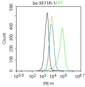
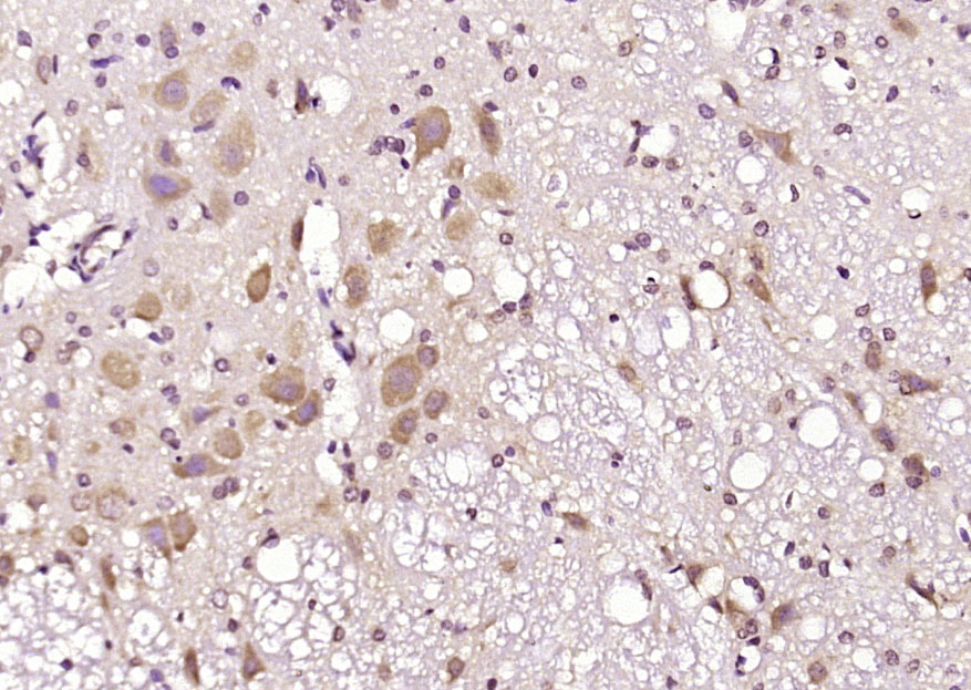
| Dilution | IHC-P=1:100-500,IHC-F=1:100-500,IF=1:100-500,Flow-Cyt=1ug/Test |
| Format | 0.01M TBS(pH7.4) with 1% BSA, 0.09% (W/V) sodium azide and 50% Glyce |
| Storage | Store at -20 °C for one year. Avoid repeated freeze/thaw cycles. When reconstituted in sterile pH 7.4 0.01M PBS or diluent of antibody the antibody is stable for at least two weeks at 2-4 °C. |
| Name | Wipi1 |
|---|---|
| Synonyms | D11Ertd498e |
| Function | Component of the autophagy machinery that controls the major intracellular degradation process by which cytoplasmic materials are packaged into autophagosomes and delivered to lysosomes for degradation (PubMed:22275429). Plays an important role in starvation- and calcium- mediated autophagy, as well as in mitophagy. Functions downstream of the ULK1 and PI3-kinases that produce phosphatidylinositol 3-phosphate (PtdIns3P) on membranes of the endoplasmic reticulum once activated. Binds phosphatidylinositol 3-phosphate (PtdIns3P), and maybe other phosphoinositides including PtdIns3,5P2 and PtdIns5P, and is recruited to phagophore assembly sites at the endoplasmic reticulum membranes. There, it assists WIPI2 in the recruitment of ATG12-ATG5-ATG16L1, a complex that directly controls the elongation of the nascent autophagosomal membrane. Together with WDR45/WIPI4, promotes ATG2 (ATG2A or ATG2B)-mediated lipid transfer by enhancing ATG2-association with phosphatidylinositol 3-monophosphate (PI3P)-containing membranes. Involved in xenophagy of Staphylococcus aureus. Invading S.aureus cells become entrapped in autophagosome-like WIPI1 positive vesicles targeted for lysosomal degradation. Also plays a distinct role in controlling the transcription of melanogenic enzymes and melanosome maturation, a process that is distinct from starvation-induced autophagy. May also regulate the trafficking of proteins involved in the mannose-6- phosphate receptor (MPR) recycling pathway (By similarity). |
| Cellular Location | Golgi apparatus, trans-Golgi network {ECO:0000250|UniProtKB:Q5MNZ9}. Endosome {ECO:0000250|UniProtKB:Q5MNZ9}. Cytoplasmic vesicle, clathrin-coated vesicle {ECO:0000250|UniProtKB:Q5MNZ9}. Preautophagosomal structure membrane; Peripheral membrane protein. Cytoplasm, cytoskeleton {ECO:0000250|UniProtKB:Q5MNZ9}. Note=Trans elements of the Golgi and peripheral endosomes. Dynamically cycles through these compartments and is susceptible to conditions that modulate membrane flux. Enriched in clathrin-coated vesicles. Upon starvation-induced autophagy, accumulates at subcellular structures in the cytoplasm: enlarged vesicular and lasso-like structures, and large cup-shaped structures predominantly around the nucleus. Recruitment to autophagic membranes is controlled by MTMR14. Labile microtubules specifically recruit markers of autophagosome formation like WIPI1, whereas mature autophagosomes may bind to stable microtubules {ECO:0000250|UniProtKB:Q5MNZ9} |
Research Areas
For Research Use Only. Not For Use In Diagnostic Procedures.
Application Protocols
Provided below are standard protocols that you may find useful for product applications.
BACKGROUND
This product as supplied is intended for research use only, not for use in human, therapeutic or diagnostic applications.
终于等到您。ABCEPTA(百远生物)抗体产品。
点击下方“我要评价 ”按钮提交您的反馈信息,您的反馈和评价是我们最宝贵的财富之一,
我们将在1-3个工作日内处理您的反馈信息。
如有疑问,联系:0512-88856768 tech-china@abcepta.com.


 癌症的基本特征包括细胞增殖、血管生成、迁移、凋亡逃避机制和细胞永生等。找到癌症发生过程中这些通路的关键标记物和对应的抗体用于检测至关重要。
 为您推荐一个泛素化位点预测神器——泛素化分析工具,可以为您的蛋白的泛素化位点作出预测和评分。
 细胞自噬受体图形绘图工具为你的蛋白的细胞受体结合位点作出预测和评分,识别结合到自噬通路中的蛋白是非常重要的,便于让我们理解自噬在正常生理、病理过程中的作用,如发育、细胞分化、神经退化性疾病、压力条件下、感染和癌症。


