


 癌症的基本特征包括细胞增殖、血管生成、迁移、凋亡逃避机制和细胞永生等。找到癌症发生过程中这些通路的关键标记物和对应的抗体用于检测至关重要。
 为您推荐一个泛素化位点预测神器——泛素化分析工具,可以为您的蛋白的泛素化位点作出预测和评分。
 细胞自噬受体图形绘图工具为你的蛋白的细胞受体结合位点作出预测和评分,识别结合到自噬通路中的蛋白是非常重要的,便于让我们理解自噬在正常生理、病理过程中的作用,如发育、细胞分化、神经退化性疾病、压力条件下、感染和癌症。
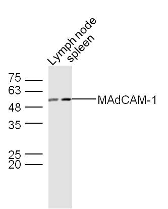
MAdCAM-1 Rabbit pAb
MAdCAM-1 Rabbit pAb

- 产品详情
- 实验流程
- 背景知识
Application
| WB, IHC-P, IHC-F, IF |
|---|---|
| Primary Accession | O70540 |
| Reactivity | Rat |
| Host | Rabbit |
| Clonality | Polyclonal |
| Calculated MW | 42507 Da |
| Physical State | Liquid |
| Immunogen | KLH conjugated synthetic peptide derived from rat MAdCAM-1 |
| Epitope Specificity | 151-250/394 |
| Isotype | IgG |
| Purity | affinity purified by Protein A |
| Buffer | 0.01M TBS (pH7.4) with 1% BSA, 0.02% Proclin300 and 50% Glycerol. |
| SUBCELLULAR LOCATION | Membrane. |
| SIMILARITY | Contains 2 Ig-like (immunoglobulin-like) domains. |
| SUBUNIT | Contains 2 Ig-like (immunoglobulin-like) domains. |
| Post-translational modifications | The Ser/Thr-rich mucin-like domain may provide possible sites for O-glycosylation (By similarity). |
| Important Note | This product as supplied is intended for research use only, not for use in human, therapeutic or diagnostic applications. |
| Background Descriptions | The recirculation of lymphocytes through different organs is thought to be regulated by adhesion molecules recognizing tissue-specific vascular addressins on the endothelium. The mucosal vascular addressin, MadCAM-1 (mucosal addressin cell adhesion molecule 1), is an immunoglobulin superfamily adhesion molecule for lymphocytes that is expressed by mucosal venules and helps direct lymphocyte traffic into Peyer’s patches and the intestinal lamina propria. MadCAM-1 acts as an endothelial cell ligand for leukocyte homing receptors L-Selectin and Integrin Alpha 4/Beta 7. MadCAM-1 is strongly expressed on inflamed portal vein/sinusoidal endothelium in autoimmune-mediated liver disease and plays a major contributory role in the progression of chronic experimental autoimmune encephalomyelitis. |
| Gene ID | 54266 |
|---|---|
| Other Names | Mucosal addressin cell adhesion molecule 1, MAdCAM-1, rMAdCAM-1, Madcam1 |
| Target/Specificity | Highly expressed on high endothelial venules (HEV) and lamina propia venules found in the small intestine, and to a lesser extent in the colon and spleen. Very low levels of expression found in pancreas and brain. Not expressed in the thymus, prostate, ovaries, testis, heart, placenta, lung, liver, skeletal muscle, kidney or peripheral blood leukocytes. |
| Dilution | WB=1:500-2000,IHC-P=1:100-500,IHC-F=1:100-500,IF=1:100-500 |
| Format | 0.01M TBS(pH7.4) with 1% BSA, 0.09% (W/V) sodium azide and 50% Glyce |
| Storage | Store at -20 °C for one year. Avoid repeated freeze/thaw cycles. When reconstituted in sterile pH 7.4 0.01M PBS or diluent of antibody the antibody is stable for at least two weeks at 2-4 °C. |
| Name | Madcam1 |
|---|---|
| Function | Cell adhesion leukocyte receptor expressed by mucosal venules, helps to direct lymphocyte traffic into mucosal tissues including the Peyer patches and the intestinal lamina propria. It can bind both the integrin alpha-4/beta-7 and L-selectin, regulating both the passage and retention of leukocytes (By similarity). |
| Cellular Location | Membrane; Single-pass type I membrane protein. |
| Tissue Location | Detected in Peyer patches and mesenteric lymph nodes but not in spleen |
Research Areas
For Research Use Only. Not For Use In Diagnostic Procedures.
Application Protocols
Provided below are standard protocols that you may find useful for product applications.
BACKGROUND
This product as supplied is intended for research use only, not for use in human, therapeutic or diagnostic applications.
终于等到您。ABCEPTA(百远生物)抗体产品。
点击下方“我要评价 ”按钮提交您的反馈信息,您的反馈和评价是我们最宝贵的财富之一,
我们将在1-3个工作日内处理您的反馈信息。
如有疑问,联系:0512-88856768 tech-china@abcepta.com.


