


 癌症的基本特征包括细胞增殖、血管生成、迁移、凋亡逃避机制和细胞永生等。找到癌症发生过程中这些通路的关键标记物和对应的抗体用于检测至关重要。
 为您推荐一个泛素化位点预测神器——泛素化分析工具,可以为您的蛋白的泛素化位点作出预测和评分。
 细胞自噬受体图形绘图工具为你的蛋白的细胞受体结合位点作出预测和评分,识别结合到自噬通路中的蛋白是非常重要的,便于让我们理解自噬在正常生理、病理过程中的作用,如发育、细胞分化、神经退化性疾病、压力条件下、感染和癌症。
Nogo R Rabbit pAb
Nogo R Rabbit pAb
- 产品详情
- 实验流程
- 背景知识
Application
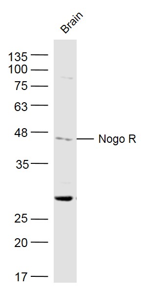
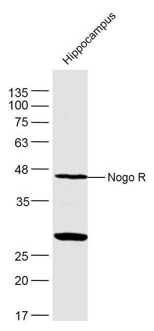
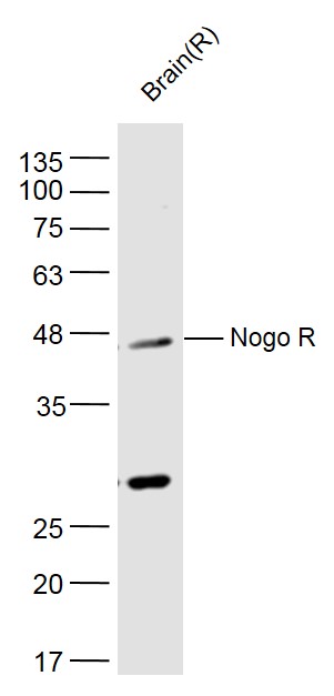
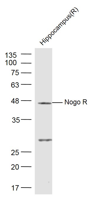
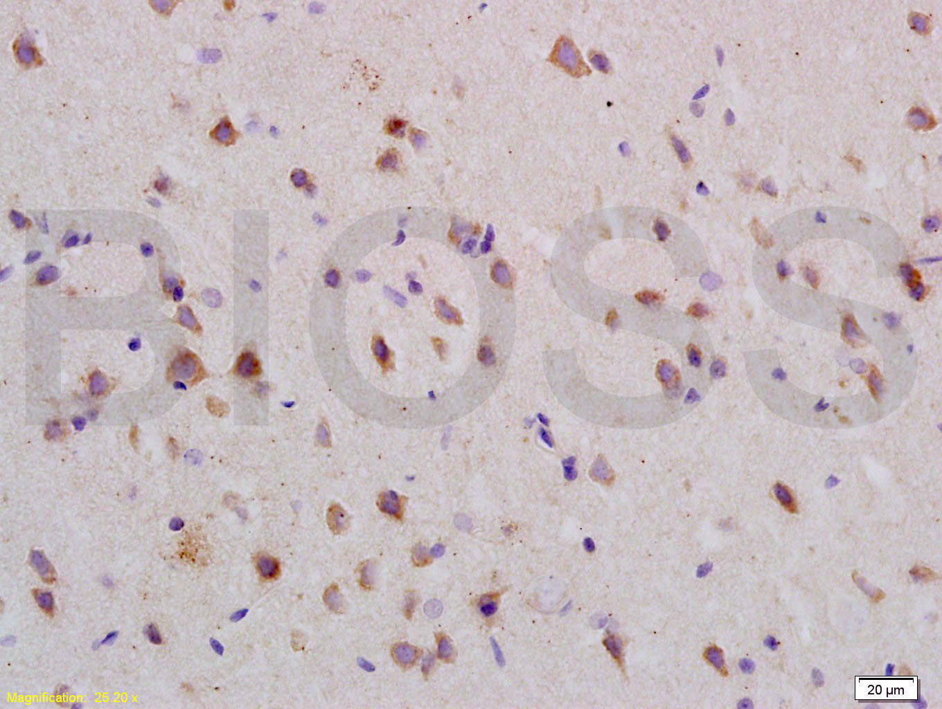
| WB, IHC-P, IHC-F, IF |
|---|---|
| Primary Accession | Q99PI8 |
| Reactivity | Mouse |
| Host | Rabbit |
| Clonality | Polyclonal |
| Calculated MW | 50987 Da |
| Physical State | Liquid |
| Immunogen | KLH conjugated synthetic peptide derived from mouse Nogo R |
| Epitope Specificity | 151-350/473 |
| Isotype | IgG |
| Purity | affinity purified by Protein A |
| Buffer | 0.01M TBS (pH7.4) with 1% BSA, 0.02% Proclin300 and 50% Glycerol. |
| SUBCELLULAR LOCATION | Cell membrane; Lipid-anchor, GPI-anchor. |
| SIMILARITY | Belongs to the Nogo receptor family. Contains 8 LRR (leucine-rich) repeats. Contains 1 LRRCT domain. Contains 1 LRRNT domain. |
| SUBUNIT | Homomultimer. Interacts with LINGO1. Interacts with KIAA0319L. |
| Important Note | This product as supplied is intended for research use only, not for use in human, therapeutic or diagnostic applications. |
| Background Descriptions | Axons are essential for neuronal communication but they do not regenerate after injury to the adult mammalian brain or spinal cord. Failed regeneration is due in part to the production of a potent axonal growth inhibitor, Nogo, by myelinating cells. The finding of a high affinity axonal receptor for the extracellular domain of Nogo provides the first insight into the basis of Nogo action. Disrupting the interaction of Nogo with the Nogo-66 receptor may facilitate axonal regeneration in vivo. The protein is dubbed the Nogo receptor because it binds with several other proteins that block neural growth. It is found to be ubiquitous in the brain and spinal cord. |
| Gene ID | 65079 |
|---|---|
| Other Names | Reticulon-4 receptor, Nogo receptor, NgR, Nogo-66 receptor, Nogo66 receptor-1, NgR1, Rtn4r, Ngr1, Nogor |
| Dilution | WB=1:500-2000,IHC-P=1:100-500,IHC-F=1:100-500,IF=1:100-500 |
| Format | 0.01M TBS(pH7.4) with 1% BSA, 0.09% (W/V) sodium azide and 50% Glyce |
| Storage | Store at -20 °C for one year. Avoid repeated freeze/thaw cycles. When reconstituted in sterile pH 7.4 0.01M PBS or diluent of antibody the antibody is stable for at least two weeks at 2-4 °C. |
| Name | Rtn4r |
|---|---|
| Synonyms | Ngr1, Nogor |
| Function | Receptor for RTN4, OMG and MAG (PubMed:11201742, PubMed:12089450, PubMed:15504325, PubMed:18411262, PubMed:22923615). Functions as a receptor for the sialylated gangliosides GT1b and GM1 (PubMed:18411262). Besides, functions as a receptor for chondroitin sulfate proteoglycans (PubMed:22406547). Can also bind heparin (PubMed:22406547). Intracellular signaling cascades are triggered via the coreceptor NGFR (By similarity). Signaling mediates activation of Rho and downstream reorganization of the actin cytoskeleton (PubMed:22325200). Mediates axonal growth inhibition (By similarity). Mediates axonal growth inhibition and plays a role in regulating axon regeneration and neuronal plasticity in the adult central nervous system (PubMed:11201742, PubMed:12089450, PubMed:15504325, PubMed:22923615). Plays a role in postnatal brain development (PubMed:27339102). Required for normal axon migration across the brain midline and normal formation of the corpus callosum (PubMed:27339102). Protects motoneurons against apoptosis; protection against apoptosis is probably mediated via interaction with MAG (PubMed:26335717). Acts in conjunction with RTN4 and LINGO1 in regulating neuronal precursor cell motility during cortical development (PubMed:20093372). Like other family members, plays a role in restricting the number dendritic spines and the number of synapses that are formed during brain development (PubMed:22325200). |
| Cellular Location | Cell membrane; Lipid-anchor, GPI-anchor. Membrane raft {ECO:0000250|UniProtKB:Q9BZR6}. Cell projection, dendrite. Cell projection, axon. Perikaryon {ECO:0000250|UniProtKB:Q99M75}. Note=Detected along dendrites and axons, close to synapses, but clearly excluded from synapses |
| Tissue Location | Detected in embryonic hippocampus neurons (PubMed:22325200). Detected in brain (at protein level) (PubMed:15504325, PubMed:22406547). Detected in neurons in the neocortex, in hippocampus, dorsal thalamus, cerebellum granule cell layer and the mitral cell layer in the olfactory bulb (PubMed:15647357). Detected in brain, dorsal root ganglion and heart |
Research Areas
For Research Use Only. Not For Use In Diagnostic Procedures.
Application Protocols
Provided below are standard protocols that you may find useful for product applications.
BACKGROUND
This product as supplied is intended for research use only, not for use in human, therapeutic or diagnostic applications.
终于等到您。ABCEPTA(百远生物)抗体产品。
点击下方“我要评价 ”按钮提交您的反馈信息,您的反馈和评价是我们最宝贵的财富之一,
我们将在1-3个工作日内处理您的反馈信息。
如有疑问,联系:0512-88856768 tech-china@abcepta.com.


