


 癌症的基本特征包括细胞增殖、血管生成、迁移、凋亡逃避机制和细胞永生等。找到癌症发生过程中这些通路的关键标记物和对应的抗体用于检测至关重要。
 为您推荐一个泛素化位点预测神器——泛素化分析工具,可以为您的蛋白的泛素化位点作出预测和评分。
 细胞自噬受体图形绘图工具为你的蛋白的细胞受体结合位点作出预测和评分,识别结合到自噬通路中的蛋白是非常重要的,便于让我们理解自噬在正常生理、病理过程中的作用,如发育、细胞分化、神经退化性疾病、压力条件下、感染和癌症。
PSMB8 Recombinant Mouse mAb
PSMB8 Recombinant Mouse mAb
- 产品详情
- 实验流程
- 背景知识
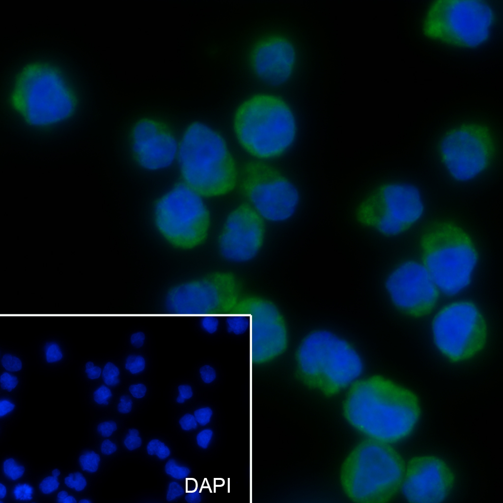
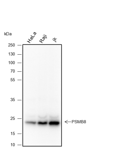
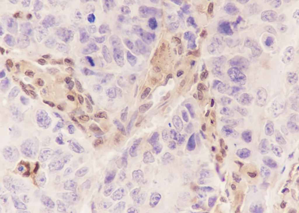
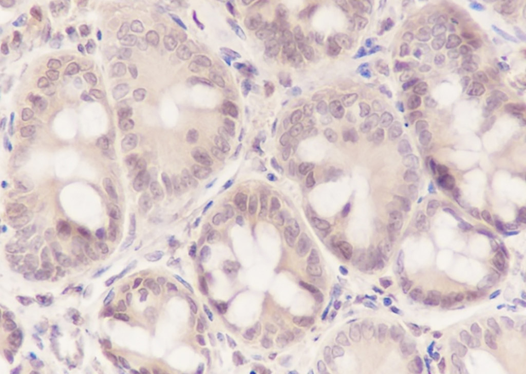
Application
| WB, IHC-P, IHC-F, IF, ICC |
|---|---|
| Host | Rabbit |
| Clonality | Recombinant |
| Physical State | Liquid |
| Isotype | IgG1, Kappa |
| Purity | affinity purified by Protein G |
| Buffer | 0.01M TBS (pH7.4) with 1% BSA, 0.02% Proclin300 and 50% Glycerol. |
| SUBCELLULAR LOCATION | Cytoplasm. Nucleus. |
| SIMILARITY | Belongs to the peptidase T1B family. |
| SUBUNIT | The 26S proteasome consists of a 20S proteasome core and two 19S regulatory subunits. The 20S proteasome core is composed of 28 subunits that are arranged in four stacked rings, resulting in a barrel-shaped structure. The two end rings are each formed by seven alpha subunits, and the two central rings are each formed by seven beta subunits. The catalytic chamber with the active sites is on the inside of the barrel. This subunit is part of the immunoproteasome where it displaces the equivalent housekeeping subunit PSMB5. Directly interacts with POMP. Interacts with HIV-1 TAT protein. Interacts with TAP1. |
| Post-translational modifications | Autocleaved. The resulting N-terminal Thr residue of the mature subunit is responsible for the nucleophile proteolytic activity. |
| DISEASE | Defects in PSMB8 are the cause of Nakajo syndrome (NKJO) [MIM:256040]; also called joint contractures muscular atrophy microcytic anemia and panniculitis-induced lipodystrophy. An autosomal recessive autoinflammatory disorder characterized by childhood onset of recurrent fever, joint stiffness and severe contractures of the hands and feet, erythematous skin lesions with subsequent development of lipodystrophy, and laboratory evidence of immune dysregulation. Accompanying features include muscle weakness and atrophy, hepatosplenomegaly, and microcytic anemia. Note=Mutation Met-75 has been found in chronic atypical neutrophilic dermatosis with lipodystrophy and elevated temperature syndrome (CANDLE syndrome). CANDLE patients have some overlapping features with NKJO patients, including a cutaneous eruption and lipodystrophy. They show a characteristic neutrophilic dermatosis with a mononuclear interstitial infiltrate in the dermis that seems pathognomonic for CANDLE syndrome (PubMed:21953331). |
| Important Note | This product as supplied is intended for research use only, not for use in human, therapeutic or diagnostic applications. |
| Background Descriptions | The proteasome is a multicatalytic proteinase complex with a highly ordered ring-shaped 20S core structure. The core structure is composed of 4 rings of 28 non-identical subunits; 2 rings are composed of 7 alpha subunits and 2 rings are composed of 7 beta subunits. Proteasomes are distributed throughout eukaryotic cells at a high concentration and cleave peptides in an ATP/ubiquitin-dependent process in a non-lysosomal pathway. An essential function of a modified proteasome, the immunoproteasome, is the processing of class I MHC peptides. This gene encodes a member of the proteasome B-type family, also known as the T1B family, that is a 20S core beta subunit. This gene is located in the class II region of the MHC (major histocompatibility complex). Expression of this gene is induced by gamma interferon and this gene product replaces catalytic subunit 3 (proteasome beta 5 subunit) in the immunoproteasome. Proteolytic processing is required to generate a mature subunit. Two alternative transcripts encoding two isoforms have been identified; both isoforms are processed to yield the same mature subunit. [provided by RefSeq, Jul 2008]. |
| Target/Specificity | Highly expressed in immature dendritic cells (at protein level). |
|---|---|
| Dilution | WB=1:500-1:1000,IHC-P=1:100-500,IHC-F=1:100-500,ICC/IF=1:50,IF=0 |
| Format | 0.01M TBS(pH7.4) with 1% BSA, 0.09% (W/V) sodium azide and 50% Glyce |
| Storage | Store at -20 °C for one year. Avoid repeated freeze/thaw cycles. When reconstituted in sterile pH 7.4 0.01M PBS or diluent of antibody the antibody is stable for at least two weeks at 2-4 °C. |
Research Areas
For Research Use Only. Not For Use In Diagnostic Procedures.
Application Protocols
Provided below are standard protocols that you may find useful for product applications.
BACKGROUND
This product as supplied is intended for research use only, not for use in human, therapeutic or diagnostic applications.
终于等到您。ABCEPTA(百远生物)抗体产品。
点击下方“我要评价 ”按钮提交您的反馈信息,您的反馈和评价是我们最宝贵的财富之一,
我们将在1-3个工作日内处理您的反馈信息。
如有疑问,联系:0512-88856768 tech-china@abcepta.com.


