


 癌症的基本特征包括细胞增殖、血管生成、迁移、凋亡逃避机制和细胞永生等。找到癌症发生过程中这些通路的关键标记物和对应的抗体用于检测至关重要。
 为您推荐一个泛素化位点预测神器——泛素化分析工具,可以为您的蛋白的泛素化位点作出预测和评分。
 细胞自噬受体图形绘图工具为你的蛋白的细胞受体结合位点作出预测和评分,识别结合到自噬通路中的蛋白是非常重要的,便于让我们理解自噬在正常生理、病理过程中的作用,如发育、细胞分化、神经退化性疾病、压力条件下、感染和癌症。
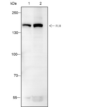
FLII Recombinant Mouse mAb
FLII Recombinant Mouse mAb


- 产品详情
- 实验流程
- 背景知识
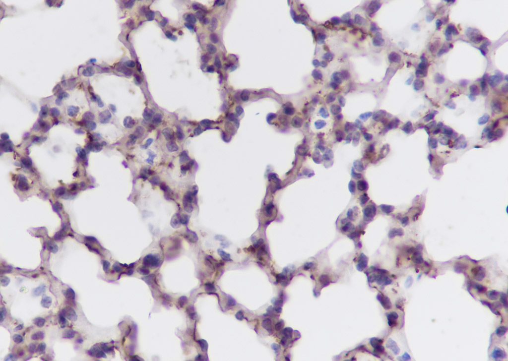
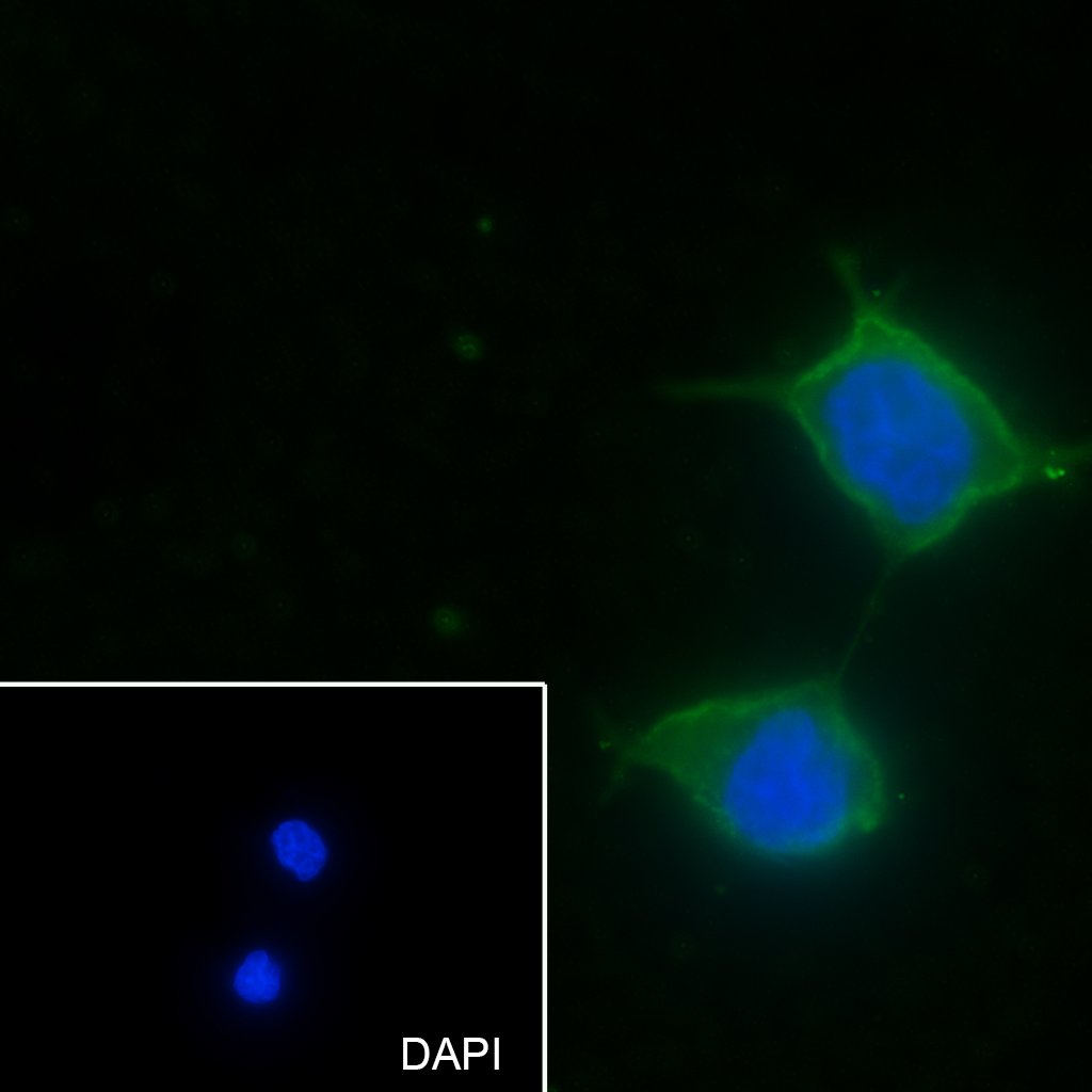
Application
| WB, IHC-P, IHC-F, IF, ICC |
|---|---|
| Host | Rabbit |
| Clonality | Recombinant |
| Physical State | Liquid |
| Isotype | IgG2a, Kappa |
| Purity | affinity purified by Protein G |
| Buffer | 0.01M TBS (pH7.4) with 1% BSA, 0.02% Proclin300 and 50% Glycerol. |
| SUBCELLULAR LOCATION | Nucleus. |
| SIMILARITY | Belongs to the ETS family. Contains 1 ETS DNA-binding domain. Contains 1 PNT (pointed) domain. |
| SUBUNIT | Can form homodimers or heterodimers with ETV6/TEL1. |
| DISEASE | Defects in FLI1 are a cause of Ewing sarcoma (ES) [MIM:612219]. A highly malignant, metastatic, primitive small round cell tumor of bone and soft tissue that affects children and adolescents. It belongs to the Ewing sarcoma family of tumors, a group of morphologically heterogeneous neoplasms that share the same cytogenetic features. They are considered neural tumors derived from cells of the neural crest. Ewing sarcoma represents the less differentiated form of the tumors. Note=A chromosomal aberration involving FLI1 is found in patients with Erwing sarcoma. Translocation t(11;22)(q24;q12) with EWSR1. |
| Important Note | This product as supplied is intended for research use only, not for use in human, therapeutic or diagnostic applications. |
| Background Descriptions | This gene encodes a protein with a gelsolin-like actin binding domain and an N-terminal leucine-rich repeat-protein protein interaction domain. The protein is similar to a Drosophila protein involved in early embryogenesis and the structural organization of indirect flight muscle. The gene is located within the Smith-Magenis syndrome region on chromosome 17. Mutations in this gene leads to abnormal muscle function, arrested development and embryonic lethality. The protein sequence shows that this might be a regulator of cytoskeleton and may have a role during cell division. |
| Dilution | WB=1:500-1:2000,IHC-P=1:100-500,IHC-F=1:100-500,ICC/IF=1:50-1:200,IF=0 |
|---|---|
| Format | 0.01M TBS(pH7.4) with 1% BSA, 0.09% (W/V) sodium azide and 50% Glyce |
| Storage | Store at -20 °C for one year. Avoid repeated freeze/thaw cycles. When reconstituted in sterile pH 7.4 0.01M PBS or diluent of antibody the antibody is stable for at least two weeks at 2-4 °C. |
Research Areas
For Research Use Only. Not For Use In Diagnostic Procedures.
Application Protocols
Provided below are standard protocols that you may find useful for product applications.
BACKGROUND
This product as supplied is intended for research use only, not for use in human, therapeutic or diagnostic applications.
终于等到您。ABCEPTA(百远生物)抗体产品。
点击下方“我要评价 ”按钮提交您的反馈信息,您的反馈和评价是我们最宝贵的财富之一,
我们将在1-3个工作日内处理您的反馈信息。
如有疑问,联系:0512-88856768 tech-china@abcepta.com.


