


 癌症的基本特征包括细胞增殖、血管生成、迁移、凋亡逃避机制和细胞永生等。找到癌症发生过程中这些通路的关键标记物和对应的抗体用于检测至关重要。
 为您推荐一个泛素化位点预测神器——泛素化分析工具,可以为您的蛋白的泛素化位点作出预测和评分。
 细胞自噬受体图形绘图工具为你的蛋白的细胞受体结合位点作出预测和评分,识别结合到自噬通路中的蛋白是非常重要的,便于让我们理解自噬在正常生理、病理过程中的作用,如发育、细胞分化、神经退化性疾病、压力条件下、感染和癌症。
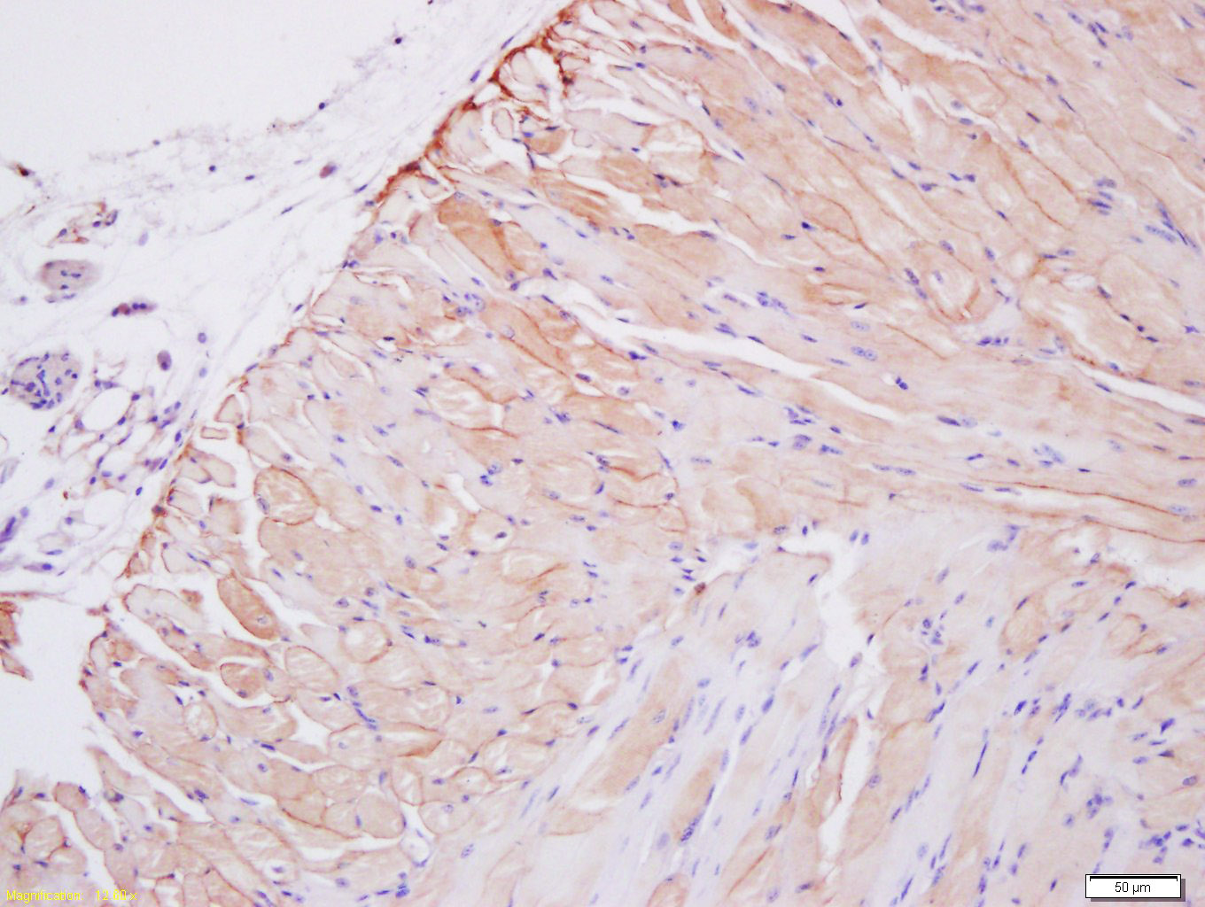
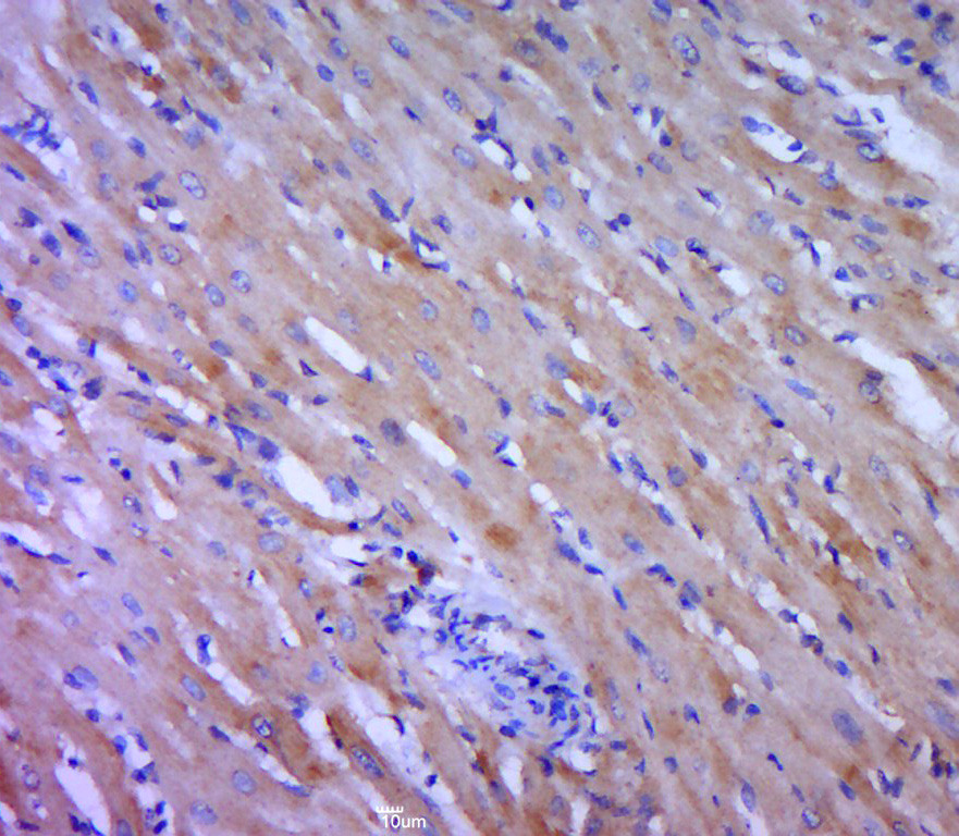
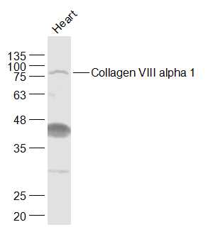
Collagen VIII alpha 1 Rabbit pAb
Collagen VIII alpha 1 Rabbit pAb
- 产品详情
- 实验流程
- 背景知识
Application
| WB, IHC-P, IHC-F, IF |
|---|---|
| Reactivity | Human |
| Host | Rabbit |
| Clonality | Polyclonal |
| Calculated MW | 19 KDa |
| Physical State | Liquid |
| Immunogen | KLH conjugated synthetic peptide derived from human Collagen VIII alpha 1 |
| Epitope Specificity | 641-744/744 |
| Isotype | IgG |
| Purity | affinity purified by Protein A |
| Buffer | 0.01M TBS (pH7.4) with 1% BSA, 0.02% Proclin300 and 50% Glycerol. |
| SUBCELLULAR LOCATION | Secreted, extracellular space, extracellular matrix, basement membrane. |
| SIMILARITY | Belongs to the PDGF/VEGF growth factor family. |
| SUBUNIT | Homodimer; antiparallel disulfide-linked dimer. Heterodimer with PDGFB; antiparallel disulfide-linked dimer. The PDGFA homodimer interacts with PDGFRA homodimers, and with heterodimers formed by PDGFRA and PDGFRB. The heterodimer composed of PDGFA and PDGFB interacts with PDGFRA homodimers, and with heterodimers formed by PDGFRA and PDGFRB. Interacts with CSPG4. |
| Post-translational modifications | Prolines at the third position of the tripeptide repeating unit (G-X-Y) are hydroxylated in some or all of the chains. Proteolytically cleaved by neutrophil elastase, in vitro. Proteolytic processing produces the C-terminal NC1 domain fragment, vastatin. |
| Important Note | This product as supplied is intended for research use only, not for use in human, therapeutic or diagnostic applications. |
| Background Descriptions | Macromolecular component of the subendothelium. Major component of the Descemet's membrane (basement membrane) of corneal endothelial cells. Also component of the endothelia of blood vessels. Necessary for migration and proliferation of vascular smooth muscle cells and thus, has a potential role in the maintenance of vessel wall integrity and structure, in particular in artherogenesis. Vastatin, the C-terminal fragment comprising the NC1 domain, inhibits aortic endothelial cell proliferation and causes cell apoptosis. |
| Target/Specificity | Expressed primarily in the subendothelium of large blood vessels. Also expressed in arterioles and venules in muscle, heart, kidney, spleen, umbilical cord, liver and lung and is also found in connective tissue layers around hair follicles, around nerve bundles in muscle, in the dura of the optic nerve, in cornea and sclera, and in the perichondrium of cartilaginous tissues. In the kidney, expressed in mesangial cells, glomerular endothelial cells, and tubular epithelial cells. Also expressed in mast cells, and in astrocytes during the repair process. Expressed in Descemet's membrane. Specifically expressed in peritoneal fibroblasts and mesothelial cells. |
|---|---|
| Dilution | WB=1:500-2000,IHC-P=1:100-500,IHC-F=1:100-500,IF=1:100-500 |
| Format | 0.01M TBS(pH7.4) with 1% BSA, 0.09% (W/V) sodium azide and 50% Glyce |
| Storage | Store at -20 °C for one year. Avoid repeated freeze/thaw cycles. When reconstituted in sterile pH 7.4 0.01M PBS or diluent of antibody the antibody is stable for at least two weeks at 2-4 °C. |
Research Areas
For Research Use Only. Not For Use In Diagnostic Procedures.
Application Protocols
Provided below are standard protocols that you may find useful for product applications.
BACKGROUND
This product as supplied is intended for research use only, not for use in human, therapeutic or diagnostic applications.
终于等到您。ABCEPTA(百远生物)抗体产品。
点击下方“我要评价 ”按钮提交您的反馈信息,您的反馈和评价是我们最宝贵的财富之一,
我们将在1-3个工作日内处理您的反馈信息。
如有疑问,联系:0512-88856768 tech-china@abcepta.com.


