


 癌症的基本特征包括细胞增殖、血管生成、迁移、凋亡逃避机制和细胞永生等。找到癌症发生过程中这些通路的关键标记物和对应的抗体用于检测至关重要。
 为您推荐一个泛素化位点预测神器——泛素化分析工具,可以为您的蛋白的泛素化位点作出预测和评分。
 细胞自噬受体图形绘图工具为你的蛋白的细胞受体结合位点作出预测和评分,识别结合到自噬通路中的蛋白是非常重要的,便于让我们理解自噬在正常生理、病理过程中的作用,如发育、细胞分化、神经退化性疾病、压力条件下、感染和癌症。
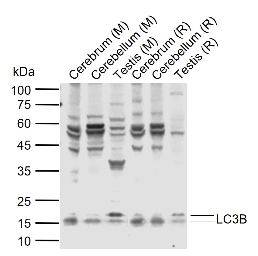
LC3B Rabbit pAb
LC3B Rabbit pAb
- 产品详情
- 实验流程
- 背景知识
Application
| WB |
|---|---|
| Primary Accession | Q9CQV6 |
| Reactivity | Mouse |
| Host | Rabbit |
| Clonality | Polyclonal |
| Calculated MW | 14617 Da |
| Physical State | Liquid |
| Immunogen | KLH conjugated synthetic peptide derived from mouse LC3B |
| Isotype | IgG |
| Purity | affinity purified by Protein A |
| Buffer | 0.01M TBS (pH7.4) with 1% BSA, 0.02% Proclin300 and 50% Glycerol. |
| SUBCELLULAR LOCATION | Cytoplasmic. Endomembrane system; Lipid-anchor. Cytoplasmic vesicle, autophagosome membrane; Lipid-anchor. Note: LC3B binds to the autophagic membranes. |
| SIMILARITY | Belongs to the MAP1 LC3 family. |
| SUBUNIT | 3 different light chains, LC1, LC2 and LC3, can associate with MAP1A and MAP1B proteins. Interacts with SQSTM1. Interacts with TP53INP1 and TP53INP2. |
| Post-translational modifications | The precursor molecule is cleaved by APG4B/ATG4B to form the cytosolic form, LC3-I. This is activated by APG7L/ATG7, transferred to ATG3 and conjugated to phospholipid to form the membrane-bound form, LC3-II. |
| Important Note | This product as supplied is intended for research use only, not for use in human, therapeutic or diagnostic applications. |
| Background Descriptions | A major contributor to cellular homeostasis is the ability of the cell to strike a balance between the formation and degradation/removal of its cellular components. This process of internal cellular turn-over is called autophagy (self-eating), and is facilitated by a pathway of around 16 interacting proteins in the human. LC3, a ubiquitin-like modifier protein, is the human homolog of yeast Apg8 and is involved in the formation of autophagosomal vacuoles, called autophagosomes. LC3 is expressed as 3 splice variants (LC3A, LC3B and LC3C), which exhibit different tissue distributions and are processed into cytosolic and autophagosomal membrane-bound forms, termed LC3-I and LC3-II, respectively. A disruption to the autophagic process is now associated with the progression of several cancers, neurodegenerative disorders and cardiac pathologies, where LC3 is widely employed as a marker for autophagy. |
| Gene ID | 67443 |
|---|---|
| Other Names | Microtubule-associated protein 1 light chain 3 beta, Autophagy-related protein LC3 B, Autophagy-related ubiquitin-like modifier LC3 B, MAP1 light chain 3-like protein 2, Microtubule-associated proteins 1A/1B light chain 3B, MAP1A/MAP1B LC3 B, MAP1A/MAP1B light chain 3 B, Map1lc3b {ECO:0000312|MGI:MGI:1914693}, Map1alc3, Map1lc3 |
| Target/Specificity | Most abundant in heart, brain, liver, skeletal muscle and testis but absent in thymus and peripheral blood leukocytes. |
| Dilution | WB=1:200-1000 |
| Format | 0.01M TBS(pH7.4) with 1% BSA, 0.09% (W/V) sodium azide and 50% Glyce |
| Storage | Store at -20 °C for one year. Avoid repeated freeze/thaw cycles. When reconstituted in sterile pH 7.4 0.01M PBS or diluent of antibody the antibody is stable for at least two weeks at 2-4 °C. |
| Name | Map1lc3b {ECO:0000312|MGI:MGI:1914693} |
|---|---|
| Synonyms | Map1alc3, Map1lc3 |
| Function | Ubiquitin-like modifier involved in formation of autophagosomal vacuoles (autophagosomes). Plays a role in mitophagy which contributes to regulate mitochondrial quantity and quality by eliminating the mitochondria to a basal level to fulfill cellular energy requirements and preventing excess ROS production. In response to cellular stress and upon mitochondria fission, binds C-18 ceramides and anchors autophagolysosomes to outer mitochondrial membranes to eliminate damaged mitochondria. While LC3s are involved in elongation of the phagophore membrane, the GABARAP/GATE-16 subfamily is essential for a later stage in autophagosome maturation. Promotes primary ciliogenesis by removing OFD1 from centriolar satellites via the autophagic pathway. Through its interaction with the reticulophagy receptor TEX264, participates in the remodeling of subdomains of the endoplasmic reticulum into autophagosomes upon nutrient stress, which then fuse with lysosomes for endoplasmic reticulum turnover. Upon nutrient stress, directly recruits cofactor JMY to the phagophore membrane surfaces and promotes JMY's actin nucleation activity and autophagosome biogenesis during autophagy. |
| Cellular Location | Cytoplasmic vesicle, autophagosome membrane; Lipid- anchor {ECO:0000250|UniProtKB:Q9GZQ8}. Endomembrane system; Lipid-anchor {ECO:0000250|UniProtKB:Q9GZQ8}. Mitochondrion membrane {ECO:0000250|UniProtKB:Q9GZQ8}; Lipid-anchor {ECO:0000250|UniProtKB:Q9GZQ8}. Cytoplasm, cytoskeleton. Cytoplasmic vesicle {ECO:0000250|UniProtKB:Q9GZQ8}. Note=LC3-II binds to the autophagic membranes. LC3-II localizes with the mitochondrial inner membrane during Parkin-mediated mitophagy (By similarity). Also localizes to discrete punctae along the ciliary axoneme (By similarity) {ECO:0000250|UniProtKB:Q9GZQ8} |
Research Areas
For Research Use Only. Not For Use In Diagnostic Procedures.
Application Protocols
Provided below are standard protocols that you may find useful for product applications.
BACKGROUND
This product as supplied is intended for research use only, not for use in human, therapeutic or diagnostic applications.
终于等到您。ABCEPTA(百远生物)抗体产品。
点击下方“我要评价 ”按钮提交您的反馈信息,您的反馈和评价是我们最宝贵的财富之一,
我们将在1-3个工作日内处理您的反馈信息。
如有疑问,联系:0512-88856768 tech-china@abcepta.com.


