


 癌症的基本特征包括细胞增殖、血管生成、迁移、凋亡逃避机制和细胞永生等。找到癌症发生过程中这些通路的关键标记物和对应的抗体用于检测至关重要。
 为您推荐一个泛素化位点预测神器——泛素化分析工具,可以为您的蛋白的泛素化位点作出预测和评分。
 细胞自噬受体图形绘图工具为你的蛋白的细胞受体结合位点作出预测和评分,识别结合到自噬通路中的蛋白是非常重要的,便于让我们理解自噬在正常生理、病理过程中的作用,如发育、细胞分化、神经退化性疾病、压力条件下、感染和癌症。
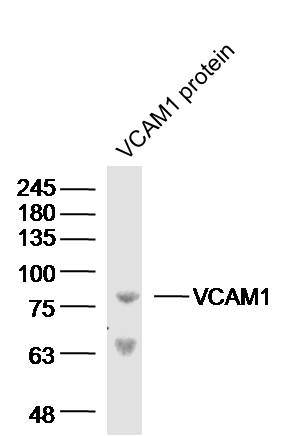
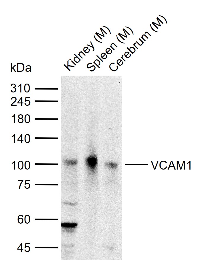
VCAM1 Rabbit pAb
VCAM1 Rabbit pAb
- 产品详情
- 实验流程
- 背景知识
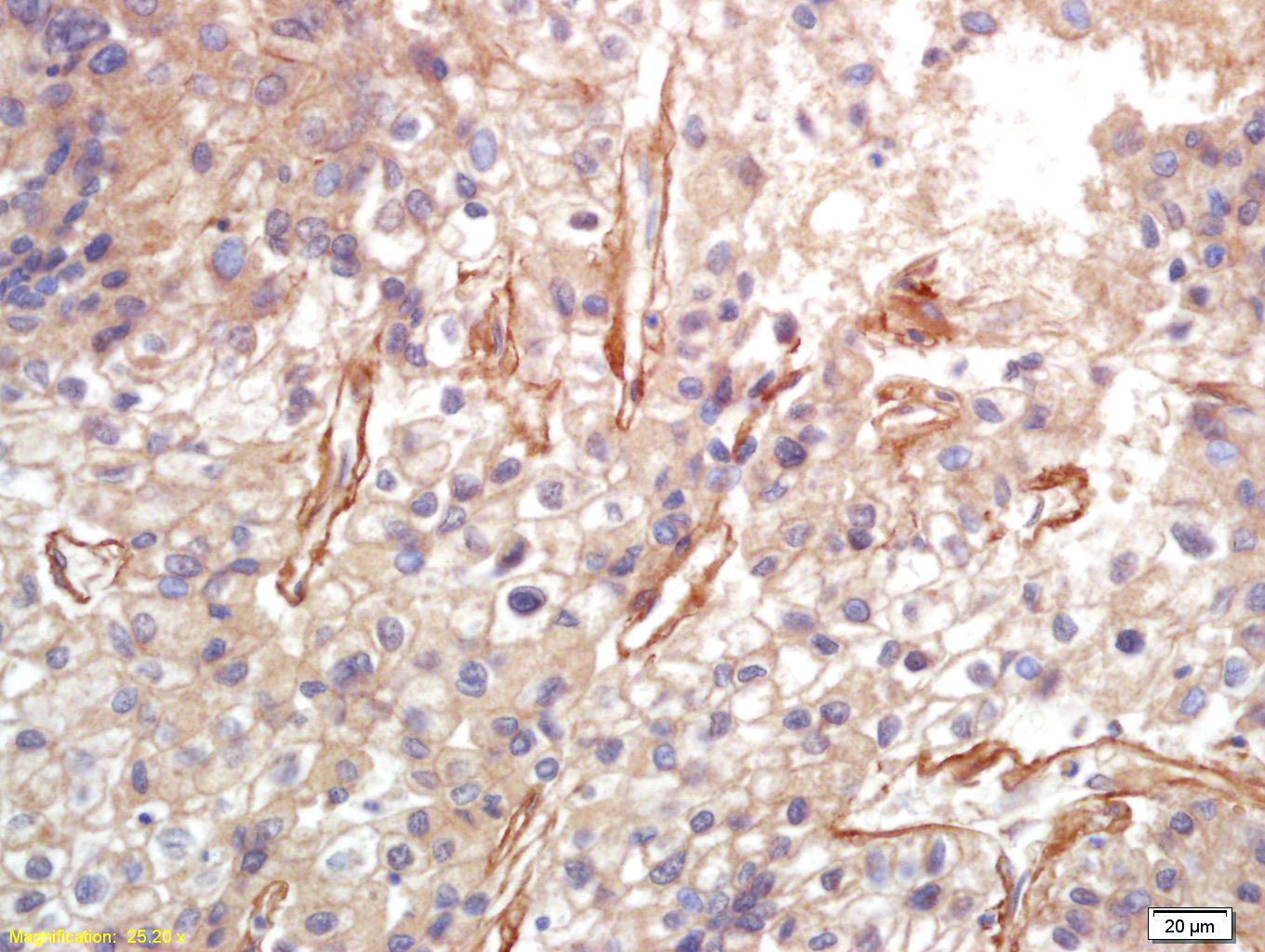
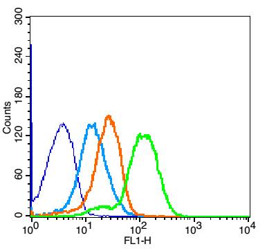
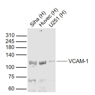
Application
| WB, IHC-P, IHC-F, IF, E |
|---|---|
| Reactivity | Mouse |
| Host | Rabbit |
| Clonality | Polyclonal |
| Calculated MW | 81 KDa |
| Physical State | Liquid |
| Immunogen | Recombinant Mouse VCAM 1 protein |
| Isotype | IgG |
| Purity | affinity purified by Protein A |
| Buffer | 0.01M TBS (pH7.4) with 1% BSA, 0.02% Proclin300 and 50% Glycerol. |
| SUBCELLULAR LOCATION | Isoform 1: Cell membrane; Single-pass type I membrane protein. Isoform 2: Cell membrane; Lipid-anchor, GPI-anchor. |
| SIMILARITY | Contains 7 Ig-like C2-type (immunoglobulin-like) domains. |
| SUBUNIT | Binds to ECMV-D capsid proteins and acts as a receptor for this virus. |
| Important Note | This product as supplied is intended for research use only, not for use in human, therapeutic or diagnostic applications. |
| Background Descriptions | VCAM1 is important in cell-cell recognition. Appears to function in leukocyte-endothelial cell adhesion. Interacts with the integrins alpha4 beta1 (beta 1 integrin VLA4) and alpha4 beta7 on leukocytes, and mediates both adhesion and signal transduction. The VCAM1/VLA4 interaction may play a pathophysiologic role both in immune responses and in leukocyte emigration to sites of inflammation. VCAM1 is also expressed by several non endothelial cell types including some macrophages, follicular dendritic cells and bone marrow, stromal cells. |
| Target/Specificity | Expressed on inflamed vascular endothelium, as well as on macrophage-like and dendritic cell types in both normal and inflamed tissue. Expressed in the bone marrow. |
|---|---|
| Dilution | WB=1:500-2000,IHC-P=1:100-500,IHC-F=1:100-500,IF=1:100-500,Flow-Cyt=1 µg/Test,ELISA=1:5000-10000 |
| Format | 0.01M TBS(pH7.4) with 1% BSA, 0.09% (W/V) sodium azide and 50% Glyce |
| Storage | Store at -20 °C for one year. Avoid repeated freeze/thaw cycles. When reconstituted in sterile pH 7.4 0.01M PBS or diluent of antibody the antibody is stable for at least two weeks at 2-4 °C. |
Research Areas
For Research Use Only. Not For Use In Diagnostic Procedures.
Application Protocols
Provided below are standard protocols that you may find useful for product applications.
BACKGROUND
This product as supplied is intended for research use only, not for use in human, therapeutic or diagnostic applications.
终于等到您。ABCEPTA(百远生物)抗体产品。
点击下方“我要评价 ”按钮提交您的反馈信息,您的反馈和评价是我们最宝贵的财富之一,
我们将在1-3个工作日内处理您的反馈信息。
如有疑问,联系:0512-88856768 tech-china@abcepta.com.


