


 癌症的基本特征包括细胞增殖、血管生成、迁移、凋亡逃避机制和细胞永生等。找到癌症发生过程中这些通路的关键标记物和对应的抗体用于检测至关重要。
 为您推荐一个泛素化位点预测神器——泛素化分析工具,可以为您的蛋白的泛素化位点作出预测和评分。
 细胞自噬受体图形绘图工具为你的蛋白的细胞受体结合位点作出预测和评分,识别结合到自噬通路中的蛋白是非常重要的,便于让我们理解自噬在正常生理、病理过程中的作用,如发育、细胞分化、神经退化性疾病、压力条件下、感染和癌症。
Mapre2 (16M10) Rat Monoclonal Antibody
Mapre2 (16M10) Rat Monoclonal Antibody
- 产品详情
- 实验流程
Application
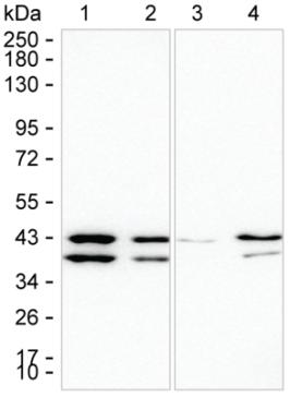
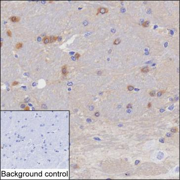
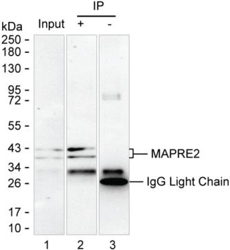
| WB, IHC, IP |
|---|---|
| Primary Accession | Q8R001 |
| Reactivity | Rat, Human, Mouse, Hamster |
| Clonality | Monoclonal |
| Calculated MW | 36946 Da |
| Gene ID | 212307 |
|---|---|
| Other Names | Microtubule-associated protein RP/EB family member 2, APC-binding protein EB2, End-binding protein 2, EB2, Mapre2 |
| Dilution | WB~~1:1000 IHC~~1:100~500 IP~~N/A |
| Storage Conditions | -20℃ |
| Name | Mapre2 |
|---|---|
| Function | Adapter protein that is involved in microtubule polymerization, and spindle function by stabilizing microtubules and anchoring them at centrosomes. Therefore, ensures mitotic progression and genome stability (By similarity). Acts as a central regulator of microtubule reorganization in apico-basal epithelial differentiation (PubMed:23813963). Plays a role during oocyte meiosis by regulating microtubule dynamics (PubMed:35398309). Participates in neurite growth by interacting with plexin B3/PLXNB3 and microtubule reorganization during apico-basal epithelial differentiation (By similarity). Plays also an essential role for cell migration and focal adhesion dynamics. Mechanistically, recruits HAX1 to microtubules in order to regulate focal adhesion dynamics (By similarity). |
| Cellular Location | Cytoplasm. Cytoplasm, cytoskeleton. Cytoplasm, cytoskeleton, spindle. Note=Associated with the microtubule network. Accumulates at the plus end of microtubules (PubMed:23813963). |
| Tissue Location | Expressed during early stages of apico-basal epithelial differentiation but down-regulated in most cells at later stages. |
Research Areas
For Research Use Only. Not For Use In Diagnostic Procedures.
Application Protocols
Provided below are standard protocols that you may find useful for product applications.
终于等到您。ABCEPTA(百远生物)抗体产品。
点击下方“我要评价 ”按钮提交您的反馈信息,您的反馈和评价是我们最宝贵的财富之一,
我们将在1-3个工作日内处理您的反馈信息。
如有疑问,联系:0512-88856768 tech-china@abcepta.com.


