


 癌症的基本特征包括细胞增殖、血管生成、迁移、凋亡逃避机制和细胞永生等。找到癌症发生过程中这些通路的关键标记物和对应的抗体用于检测至关重要。
 为您推荐一个泛素化位点预测神器——泛素化分析工具,可以为您的蛋白的泛素化位点作出预测和评分。
 细胞自噬受体图形绘图工具为你的蛋白的细胞受体结合位点作出预测和评分,识别结合到自噬通路中的蛋白是非常重要的,便于让我们理解自噬在正常生理、病理过程中的作用,如发育、细胞分化、神经退化性疾病、压力条件下、感染和癌症。
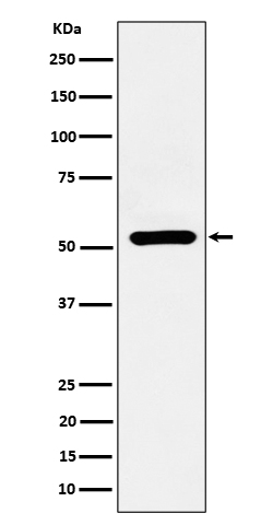
ADRA1A Antibody
Rabbit mAb
- 产品详情
- 实验流程
Application
| WB, IF, FC, ICC |
|---|---|
| Primary Accession | P35348 |
| Reactivity | Rat, Human, Mouse |
| Clonality | Monoclonal |
| Other Names | ADRA1A; Adra1c; ADRA1L1; ALPHA1AAR; |
| Isotype | Rabbit IgG |
| Host | Rabbit |
| Calculated MW | 51487 Da |
| Dilution | WB 1:500~1:2000 ICC/IF 1:50~1:200 FC 1:50 |
|---|---|
| Purification | Affinity-chromatography |
| Immunogen | A synthesized peptide derived from human ADRA1A |
| Description | This alpha-adrenergic receptor mediates its action by association with G proteins that activate a phosphatidylinositol-calcium second messenger system. Its effect is mediated by G(q) and G(11) proteins. |
| Storage Condition and Buffer | Rabbit IgG in phosphate buffered saline , pH 7.4, 150mM NaCl, 0.02% sodium azide and 50% glycerol. Store at +4°C short term. Store at -20°C long term. Avoid freeze / thaw cycle. |
| Name | ADRA1A (HGNC:277) |
|---|---|
| Synonyms | ADRA1C |
| Function | Alpha-1 adrenergic receptors are G protein-coupled receptors for catecholamines that signal through the G(q) family of G proteins, including G(q) and G(11). Upon activation, they stimulate the phosphatidylinositol-calcium second messenger pathway, leading to calcium release from intracellular stores and activation of protein kinase C (PubMed:37563160). ADRA1A binds the catecholamine ligands norepinephrine and epinephrine (PubMed:18802028, PubMed:37563160, PubMed:7815325, PubMed:8024574, PubMed:8183249, PubMed:8832064). Can also couple to G(14) protein (By similarity). Nuclear ADRA1A forms heterooligomers with ADRA1B to regulate phenylephrine(PE)-stimulated ERK signaling in cardiac myocytes (PubMed:18802028, PubMed:22120526). At the plasma membrane, ADRA1A interacts with CAVIN4/MURC to regulates ERK activation in cardiomyocytes, contributing to the regulation of cardiac hypertrophy (PubMed:24567387). Additionally, functions as a vasopressor in resistance arteries and plays a role in maintaining normal arterial blood pressure (By similarity). |
| Cellular Location | Nucleus membrane; Multi-pass membrane protein. Cell membrane; Multi-pass membrane protein. Cytoplasm. Membrane, caveola. Note=Location at the nuclear membrane facilitates heterooligomerization and regulates ERK-mediated signaling in cardiac myocytes (PubMed:18802028, PubMed:22120526) Colocalizes with GNAQ, PLCB1 as well as LAP2 at the nuclear membrane of cardiac myocytes (PubMed:18802028, PubMed:22120526). Colocalizes with CAVIN4 and CAV3 at the plasma membrane and partly within the cytoplasm in cardiomyocytes (PubMed:24567387). |
| Tissue Location | Expressed in heart, brain, liver and prostate, but not in kidney, lung, adrenal, aorta and pituitary. Within the prostate, expressed in the apex, base, periurethral and lateral lobe. Isoform 4 is the most abundant isoform expressed in the prostate with high levels also detected in liver and heart. |
Research Areas
For Research Use Only. Not For Use In Diagnostic Procedures.
Application Protocols
Provided below are standard protocols that you may find useful for product applications.
终于等到您。ABCEPTA(百远生物)抗体产品。
点击下方“我要评价 ”按钮提交您的反馈信息,您的反馈和评价是我们最宝贵的财富之一,
我们将在1-3个工作日内处理您的反馈信息。
如有疑问,联系:0512-88856768 tech-china@abcepta.com.
¥ 1,500.00
Cat# AP92800


