


 癌症的基本特征包括细胞增殖、血管生成、迁移、凋亡逃避机制和细胞永生等。找到癌症发生过程中这些通路的关键标记物和对应的抗体用于检测至关重要。
 为您推荐一个泛素化位点预测神器——泛素化分析工具,可以为您的蛋白的泛素化位点作出预测和评分。
 细胞自噬受体图形绘图工具为你的蛋白的细胞受体结合位点作出预测和评分,识别结合到自噬通路中的蛋白是非常重要的,便于让我们理解自噬在正常生理、病理过程中的作用,如发育、细胞分化、神经退化性疾病、压力条件下、感染和癌症。
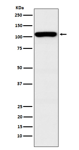
GEF H1 Antibody
Rabbit mAb
- 产品详情
- 实验流程
Application
| WB, IF, FC, ICC |
|---|---|
| Primary Accession | Q92974 |
| Reactivity | Rat, Human, Mouse |
| Clonality | Monoclonal |
| Other Names | ARHG2; ARHGEF2; GEF; GEFH1; Lbcl1; Lfc; LFP40; |
| Isotype | Rabbit IgG |
| Host | Rabbit |
| Calculated MW | 111543 Da |
| Dilution | WB 1:500~1:2000 ICC/IF 1:50~1:200 FC 1:60 |
|---|---|
| Purification | Affinity-chromatography |
| Immunogen | A synthesized peptide derived from human GEF H1 |
| Description | Activates Rho-GTPases by promoting the exchange of GDP for GTP. May be involved in epithelial barrier permeability, cell motility and polarization, dendritic spine morphology, antigen presentation, leukemic cell differentiation, cell cycle regulation, and cancer. |
| Storage Condition and Buffer | Rabbit IgG in phosphate buffered saline , pH 7.4, 150mM NaCl, 0.02% sodium azide and 50% glycerol. Store at +4°C short term. Store at -20°C long term. Avoid freeze / thaw cycle. |
| Name | ARHGEF2 |
|---|---|
| Synonyms | KIAA0651, LFP40 |
| Function | Activates Rho-GTPases by promoting the exchange of GDP for GTP. May be involved in epithelial barrier permeability, cell motility and polarization, dendritic spine morphology, antigen presentation, leukemic cell differentiation, cell cycle regulation, innate immune response, and cancer. Binds Rac-GTPases, but does not seem to promote nucleotide exchange activity toward Rac-GTPases, which was uniquely reported in PubMed:9857026. May stimulate instead the cortical activity of Rac. Inactive toward CDC42, TC10, or Ras-GTPases. Forms an intracellular sensing system along with NOD1 for the detection of microbial effectors during cell invasion by pathogens. Required for RHOA and RIP2 dependent NF-kappaB signaling pathways activation upon S.flexneri cell invasion. Involved not only in sensing peptidoglycan (PGN)-derived muropeptides through NOD1 that is independent of its GEF activity, but also in the activation of NF-kappaB by Shigella effector proteins (IpgB2 and OspB) which requires its GEF activity and the activation of RhoA. Involved in innate immune signaling transduction pathway promoting cytokine IL6/interleukin-6 and TNF secretion in macrophage upon stimulation by bacterial peptidoglycans; acts as a signaling intermediate between NOD2 receptor and RIPK2 kinase. Contributes to the tyrosine phosphorylation of RIPK2 through Src tyrosine kinase leading to NF-kappaB activation by NOD2. Overexpression activates Rho-, but not Rac-GTPases, and increases paracellular permeability (By similarity). Involved in neuronal progenitor cell division and differentiation (PubMed:28453519). Involved in the migration of precerebellar neurons (By similarity). |
| Cellular Location | Cytoplasm, cytoskeleton. Cytoplasm. Cell junction, tight junction. Golgi apparatus. Cytoplasm, cytoskeleton, spindle. Cell projection, ruffle membrane. Cytoplasmic vesicle. Note=Localizes to the tips of cortical microtubules of the mitotic spindle during cell division, and is further released upon microtubule depolymerization (PubMed:15827085) Recruited into membrane ruffles induced by S.flexneri at tight junctions of polarized epithelial cells (PubMed:19043560). Colocalized with NOD2 and RIPK2 in vesicles and with the cytoskeleton (PubMed:21887730). |
Research Areas
For Research Use Only. Not For Use In Diagnostic Procedures.
Application Protocols
Provided below are standard protocols that you may find useful for product applications.
终于等到您。ABCEPTA(百远生物)抗体产品。
点击下方“我要评价 ”按钮提交您的反馈信息,您的反馈和评价是我们最宝贵的财富之一,
我们将在1-3个工作日内处理您的反馈信息。
如有疑问,联系:0512-88856768 tech-china@abcepta.com.
¥ 1,500.00
Cat# AP91820


