


 癌症的基本特征包括细胞增殖、血管生成、迁移、凋亡逃避机制和细胞永生等。找到癌症发生过程中这些通路的关键标记物和对应的抗体用于检测至关重要。
 为您推荐一个泛素化位点预测神器——泛素化分析工具,可以为您的蛋白的泛素化位点作出预测和评分。
 细胞自噬受体图形绘图工具为你的蛋白的细胞受体结合位点作出预测和评分,识别结合到自噬通路中的蛋白是非常重要的,便于让我们理解自噬在正常生理、病理过程中的作用,如发育、细胞分化、神经退化性疾病、压力条件下、感染和癌症。
Histone H1.0 Antibody
Rabbit mAb


- 产品详情
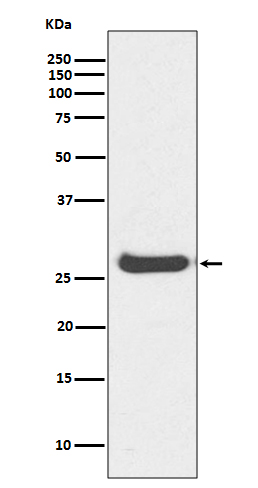
- 实验流程
Application
| WB, IHC, IF, ICC, IHF |
|---|---|
| Primary Accession | P07305 |
| Reactivity | Rat, Human, Mouse |
| Clonality | Monoclonal |
| Other Names | Histone H1.0; Histone H1(0); Histone H1.0, N-terminally processed; H1F0; H1FV; Histone H5; |
| Isotype | Rabbit IgG |
| Host | Rabbit |
| Calculated MW | 20863 Da |
| Dilution | WB 1:1000~1:2000 IHC 1:50~1:200 ICC/IF 1:50~1:200 |
|---|---|
| Purification | Affinity-chromatography |
| Immunogen | A synthesized peptide derived from human Histone H1.0 |
| Description | Histone H1.0 is a lysine rich member of the H1 family of linker histones. The H1 family of proteins interacts with linker DNA between nucleosomes and mediates compaction into higher order chromatin. Histones H1 are necessary for the condensation of nucleosome chains into higher-order structures. The H1F0 histones are found in cells that are in terminal stages of differentiation or that have low rates of cell division. |
| Storage Condition and Buffer | Rabbit IgG in phosphate buffered saline , pH 7.4, 150mM NaCl, 0.02% sodium azide and 50% glycerol. Store at +4°C short term. Store at -20°C long term. Avoid freeze / thaw cycle. |
| Name | H1-0 (HGNC:4714) |
|---|---|
| Function | Histone H1 protein binds to linker DNA between nucleosomes forming the macromolecular structure known as the chromatin fiber (PubMed:33238161). Histones H1 are necessary for the condensation of nucleosome chains into higher-order structured fibers and promote formation of the H3K27me3 mark by the PRC2/EED-EZH2 complex (PubMed:33238161). The histones H1.0 are found in cells that are in terminal stages of differentiation or that have low rates of cell division (PubMed:7374750). |
| Cellular Location | Nucleus {ECO:0000255|PROSITE-ProRule:PRU00837, ECO:0000269|PubMed:18993075}. Nucleus, nucleolus. Chromosome {ECO:0000255|PROSITE- ProRule:PRU00837, ECO:0000269|PubMed:18993075, ECO:0000269|PubMed:25645921, ECO:0000269|PubMed:38530350} Note=Enriched at nucleolus-associated DNA repeats and chromatin domains (PubMed:25645921). During mitosis, it appears in the vicinity of condensed chromosomes (PubMed:18993075). The RNA edited version has been localized to nuclear speckles (PubMed:18993075). Comparted to other H1 histones, shows a less pronounced peripheral nuclear localization (PubMed:38530350). |
Research Areas
For Research Use Only. Not For Use In Diagnostic Procedures.
Application Protocols
Provided below are standard protocols that you may find useful for product applications.
终于等到您。ABCEPTA(百远生物)抗体产品。
点击下方“我要评价 ”按钮提交您的反馈信息,您的反馈和评价是我们最宝贵的财富之一,
我们将在1-3个工作日内处理您的反馈信息。
如有疑问,联系:0512-88856768 tech-china@abcepta.com.
¥ 1,500.00
Cat# AP90851


