


 癌症的基本特征包括细胞增殖、血管生成、迁移、凋亡逃避机制和细胞永生等。找到癌症发生过程中这些通路的关键标记物和对应的抗体用于检测至关重要。
 为您推荐一个泛素化位点预测神器——泛素化分析工具,可以为您的蛋白的泛素化位点作出预测和评分。
 细胞自噬受体图形绘图工具为你的蛋白的细胞受体结合位点作出预测和评分,识别结合到自噬通路中的蛋白是非常重要的,便于让我们理解自噬在正常生理、病理过程中的作用,如发育、细胞分化、神经退化性疾病、压力条件下、感染和癌症。
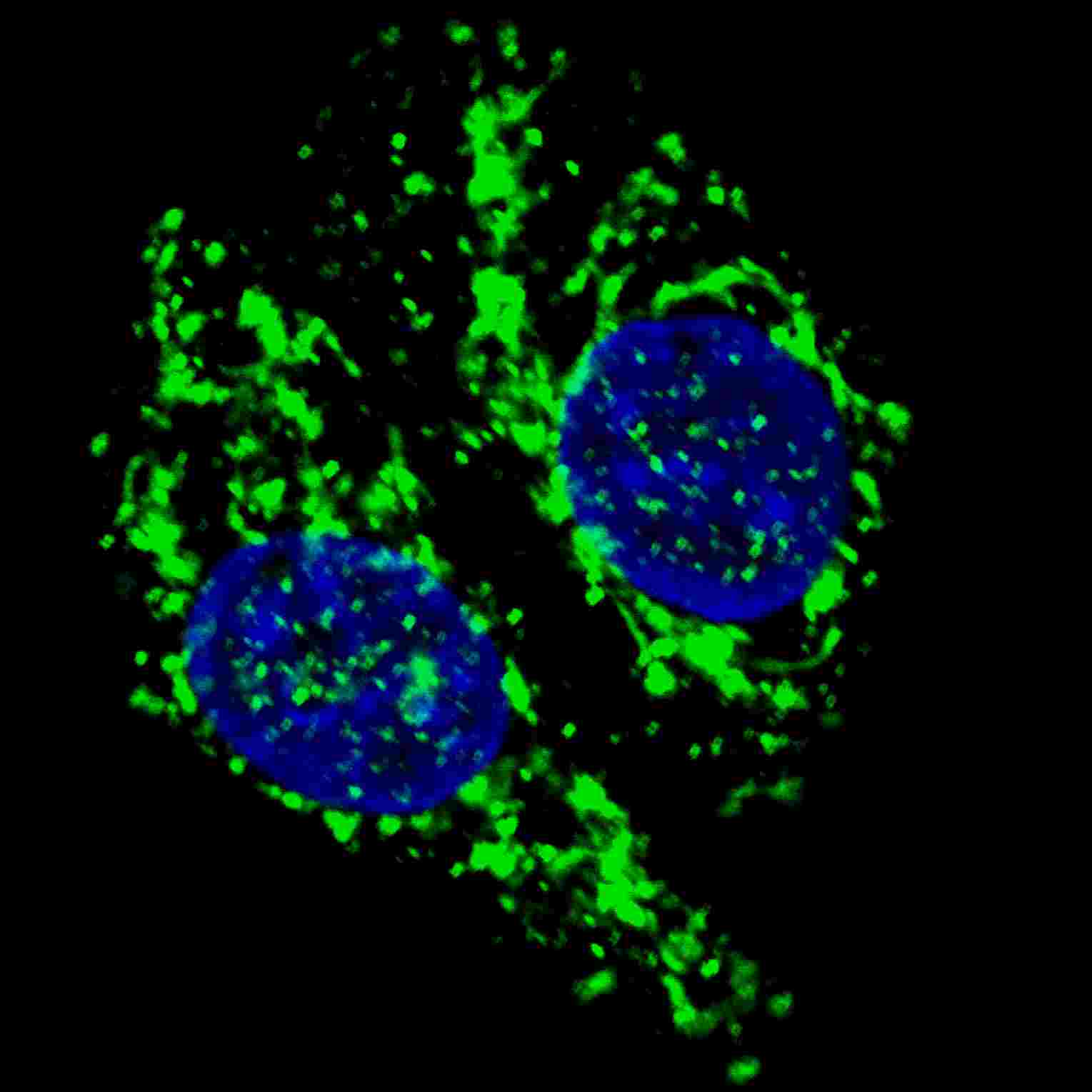
AIFM1 Antibody (N-term)
Affinity Purified Rabbit Polyclonal Antibody (Pab)
_-_U251_30.jpg)
- 产品详情
- 实验流程
- 背景知识
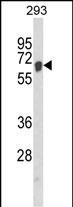
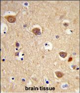
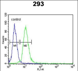
Application
| IF, WB, IHC-P, FC, E |
|---|---|
| Primary Accession | O95831 |
| Reactivity | Human |
| Host | Rabbit |
| Clonality | Polyclonal |
| Isotype | Rabbit IgG |
| Calculated MW | 66901 Da |
| Antigen Region | 70-98 aa |
| Gene ID | 9131 |
|---|---|
| Other Names | Apoptosis-inducing factor 1, mitochondrial, 111-, Programmed cell death protein 8, AIFM1, AIF, PDCD8 |
| Target/Specificity | This AIFM1 antibody is generated from rabbits immunized with a KLH conjugated synthetic peptide between 70-98 amino acids from the N-terminal region of human AIFM1. |
| Dilution | IF~~1:200 WB~~1:1000 IHC-P~~1:100~500 FC~~1:10~50 E~~Use at an assay dependent concentration. |
| Format | Purified polyclonal antibody supplied in PBS with 0.09% (W/V) sodium azide. This antibody is purified through a protein A column, followed by peptide affinity purification. |
| Storage | Maintain refrigerated at 2-8°C for up to 2 weeks. For long term storage store at -20°C in small aliquots to prevent freeze-thaw cycles. |
| Precautions | AIFM1 Antibody (N-term) is for research use only and not for use in diagnostic or therapeutic procedures. |
| Name | AIFM1 (HGNC:8768) |
|---|---|
| Synonyms | AIF, PDCD8 |
| Function | Functions both as NADH oxidoreductase and as regulator of apoptosis (PubMed:17094969, PubMed:20362274, PubMed:23217327, PubMed:33168626). In response to apoptotic stimuli, it is released from the mitochondrion intermembrane space into the cytosol and to the nucleus, where it functions as a proapoptotic factor in a caspase- independent pathway (PubMed:20362274). Release into the cytoplasm is mediated upon binding to poly-ADP-ribose chains (By similarity). The soluble form (AIFsol) found in the nucleus induces 'parthanatos' i.e. caspase-independent fragmentation of chromosomal DNA (PubMed:20362274). Binds to DNA in a sequence-independent manner (PubMed:27178839). Interacts with EIF3G, and thereby inhibits the EIF3 machinery and protein synthesis, and activates caspase-7 to amplify apoptosis (PubMed:17094969). Plays a critical role in caspase-independent, pyknotic cell death in hydrogen peroxide-exposed cells (PubMed:19418225). In contrast, participates in normal mitochondrial metabolism. Plays an important role in the regulation of respiratory chain biogenesis by interacting with CHCHD4 and controlling CHCHD4 mitochondrial import (PubMed:26004228). |
| Cellular Location | Mitochondrion intermembrane space. Mitochondrion inner membrane. Cytoplasm. Nucleus. Cytoplasm, perinuclear region. Note=Proteolytic cleavage during or just after translocation into the mitochondrial intermembrane space (IMS) results in the formation of an inner-membrane-anchored mature form (AIFmit). During apoptosis, further proteolytic processing leads to a mature form, which is confined to the mitochondrial IMS in a soluble form (AIFsol). AIFsol is released to the cytoplasm in response to specific death signals, and translocated to the nucleus, where it induces nuclear apoptosis (PubMed:15775970). Release into the cytoplasm is mediated upon binding to poly-ADP-ribose chains (By similarity) Translocation into the nucleus is promoted by interaction with (auto- poly-ADP-ribosylated) processed form of PARP1 (PubMed:33168626) Colocalizes with EIF3G in the nucleus and perinuclear region (PubMed:17094969). {ECO:0000250|UniProtKB:Q9Z0X1, ECO:0000269|PubMed:15775970, ECO:0000269|PubMed:17094969, ECO:0000269|PubMed:33168626} [Isoform 4]: Mitochondrion. Cytoplasm, cytosol. Note=In pro-apoptotic conditions, is released from mitochondria to cytosol in a calpain/cathepsin-dependent manner. |
| Tissue Location | Expressed in all tested tissues (PubMed:16644725). Detected in muscle and skin fibroblasts (at protein level) (PubMed:23217327). Expressed in osteoblasts (at protein level) (PubMed:28842795). [Isoform 4]: Expressed in all tested tissues except brain. |
For Research Use Only. Not For Use In Diagnostic Procedures.
Provided below are standard protocols that you may find useful for product applications.
BACKGROUND
AIFM1 is a flavoprotein essential for nuclear disassembly in apoptotic cells that is found in the mitochondrial intermembrane space in healthy cells. Induction of apoptosis results in the translocation of this protein to the nucleus where it effects chromosome condensation and fragmentation. In addition, this protein induces mitochondria to release the apoptogenic proteins cytochrome c and caspase-9.
REFERENCES
References for protein:
1.Daugas,E., et.al., FASEB J. 14 (5), 729-739 (2000)
2.Schulthess,F.T., et.al., PLoS ONE 4 (2), E4394 (2009)
References for U251 cell line:
1. Westermark B.; Pontén J.; Hugosson R. (1973).” Determinants for the establishment of permanent tissue culture lines from human gliomas”. Acta Pathol Microbiol Scand A. 81:791-805. [PMID: 4359449].
2. Pontén, J.,Westermark B. (1978).” Properties of Human Malignant Glioma Cells in Vitro”. Medical Biology 56: 184-193.[PMID: 359950].
3. Geng Y.;Kohli L.; Klocke B.J.; Roth K.A.(2010). “Chloroquine-induced autophagic vacuole accumulation and cell death in glioma cells is p53 independent”. Neuro Oncol. 12(5): 473–481.[ PMID: 20406898].
终于等到您。ABCEPTA(百远生物)抗体产品。
点击下方“我要评价 ”按钮提交您的反馈信息,您的反馈和评价是我们最宝贵的财富之一,
我们将在1-3个工作日内处理您的反馈信息。
如有疑问,联系:0512-88856768 tech-china@abcepta.com.


