


 癌症的基本特征包括细胞增殖、血管生成、迁移、凋亡逃避机制和细胞永生等。找到癌症发生过程中这些通路的关键标记物和对应的抗体用于检测至关重要。
 为您推荐一个泛素化位点预测神器——泛素化分析工具,可以为您的蛋白的泛素化位点作出预测和评分。
 细胞自噬受体图形绘图工具为你的蛋白的细胞受体结合位点作出预测和评分,识别结合到自噬通路中的蛋白是非常重要的,便于让我们理解自噬在正常生理、病理过程中的作用,如发育、细胞分化、神经退化性疾病、压力条件下、感染和癌症。
ATP5F1A Rabbit mAb
- 产品详情
- 实验流程
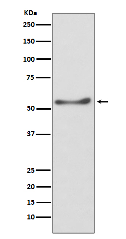
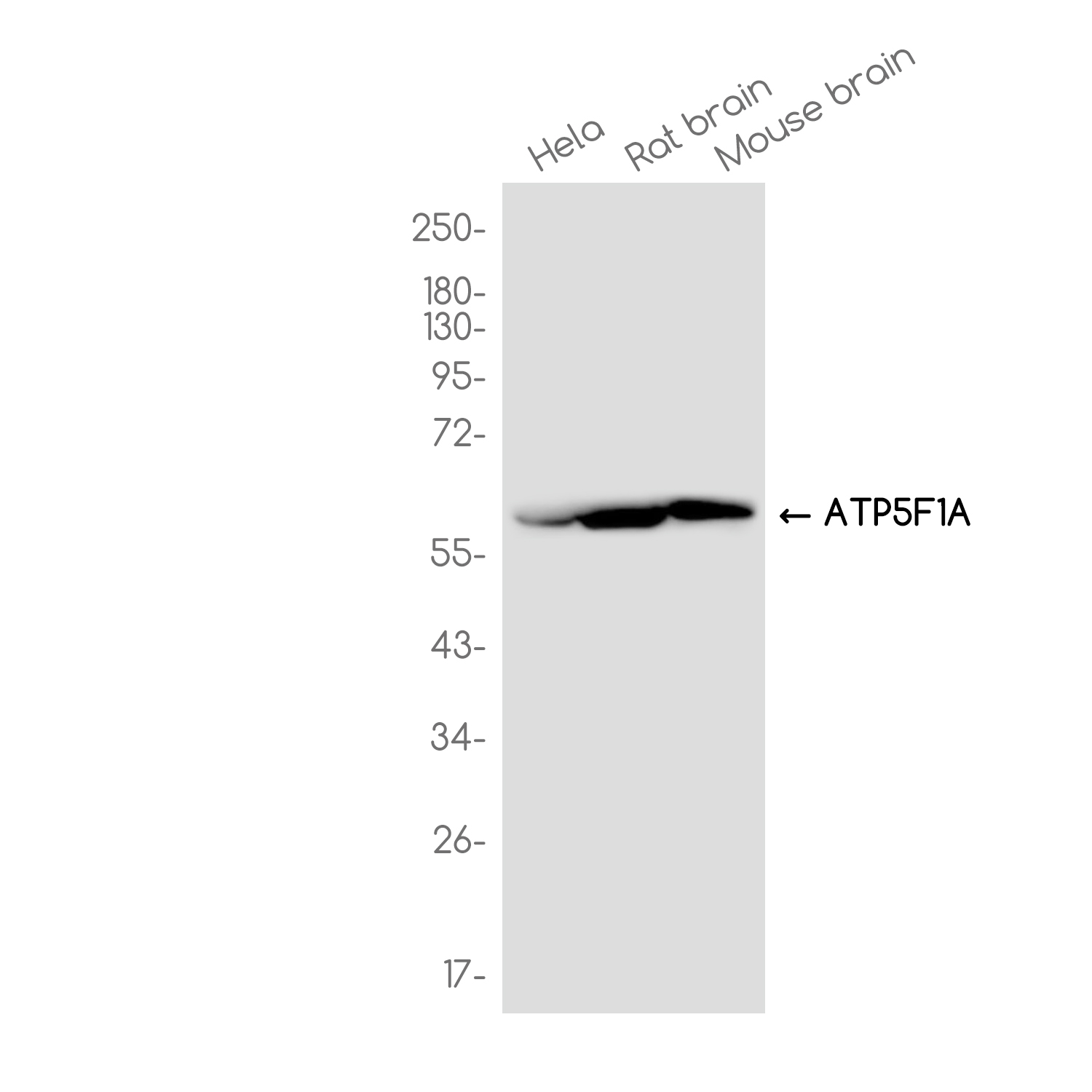
Application
| WB, IHC-P, FC, ICC |
|---|---|
| Primary Accession | P25705 |
| Reactivity | Human, Mouse, Rat |
| Host | Rabbit |
| Clonality | Monoclonal Antibody |
| Calculated MW | 59751 Da |
| Gene ID | 498 |
|---|---|
| Other Names | ATP5F1A |
| Dilution | WB~~1/500-1/1000 IHC-P~~1:50~200 FC~~1:10~50 ICC~~N/A |
| Format | 10mM PBS, pH 7.4, 150mM sodium chloride, 0.05% BSA, 0.02% sodium azide and 50% glycerol. |
| Storage | Store at 4°C short term. Aliquot and store at -20°C long term. Avoid freeze/thaw cycles. |
| Name | ATP5F1A (HGNC:823) |
|---|---|
| Function | Subunit alpha, of the mitochondrial membrane ATP synthase complex (F(1)F(0) ATP synthase or Complex V) that produces ATP from ADP in the presence of a proton gradient across the membrane which is generated by electron transport complexes of the respiratory chain (Probable). ATP synthase complex consist of a soluble F(1) head domain - the catalytic core - and a membrane F(1) domain - the membrane proton channel (PubMed:37244256). These two domains are linked by a central stalk rotating inside the F(1) region and a stationary peripheral stalk (PubMed:37244256). During catalysis, ATP synthesis in the catalytic domain of F(1) is coupled via a rotary mechanism of the central stalk subunits to proton translocation (Probable). In vivo, can only synthesize ATP although its ATP hydrolase activity can be activated artificially in vitro (By similarity). With the catalytic subunit beta (ATP5F1B), forms the catalytic core in the F(1) domain (PubMed:37244256). Subunit alpha does not bear the catalytic high- affinity ATP-binding sites (Probable). Binds the bacterial siderophore enterobactin and can promote mitochondrial accumulation of enterobactin-derived iron ions (PubMed:30146159). |
| Cellular Location | Mitochondrion. Mitochondrion inner membrane {ECO:0000250|UniProtKB:P19483}; Peripheral membrane protein {ECO:0000250|UniProtKB:P19483}; Matrix side {ECO:0000250|UniProtKB:P19483}. Cell membrane; Peripheral membrane protein; Extracellular side. Note=Colocalizes with HRG on the cell surface of T-cells (PubMed:19285951). |
| Tissue Location | Fetal lung, heart, liver, gut and kidney. Expressed at higher levels in the fetal brain, retina and spinal cord |
Research Areas
For Research Use Only. Not For Use In Diagnostic Procedures.
Application Protocols
Provided below are standard protocols that you may find useful for product applications.
终于等到您。ABCEPTA(百远生物)抗体产品。
点击下方“我要评价 ”按钮提交您的反馈信息,您的反馈和评价是我们最宝贵的财富之一,
我们将在1-3个工作日内处理您的反馈信息。
如有疑问,联系:0512-88856768 tech-china@abcepta.com.
¥ 1,500.00
Cat# AP79034


