


 癌症的基本特征包括细胞增殖、血管生成、迁移、凋亡逃避机制和细胞永生等。找到癌症发生过程中这些通路的关键标记物和对应的抗体用于检测至关重要。
 为您推荐一个泛素化位点预测神器——泛素化分析工具,可以为您的蛋白的泛素化位点作出预测和评分。
 细胞自噬受体图形绘图工具为你的蛋白的细胞受体结合位点作出预测和评分,识别结合到自噬通路中的蛋白是非常重要的,便于让我们理解自噬在正常生理、病理过程中的作用,如发育、细胞分化、神经退化性疾病、压力条件下、感染和癌症。
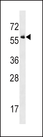
Activin Receptor Type IA (ACVR1) Antibody (Center R147)
Purified Rabbit Polyclonal Antibody (Pab)


- 产品详情
- 文献引用 : 2
- 实验流程
- 背景知识
Application
| WB, IHC-P, E |
|---|---|
| Primary Accession | Q04771 |
| Other Accession | P80201, P37172, Q28041 |
| Reactivity | Human, Rat, Mouse |
| Predicted | Bovine, Rat |
| Host | Rabbit |
| Clonality | Polyclonal |
| Isotype | Rabbit IgG |
| Calculated MW | 57153 Da |
| Antigen Region | 132-162 aa |
| Gene ID | 90 |
|---|---|
| Other Names | Activin receptor type-1, Activin receptor type I, ACTR-I, Activin receptor-like kinase 2, ALK-2, Serine/threonine-protein kinase receptor R1, SKR1, TGF-B superfamily receptor type I, TSR-I, ACVR1, ACVRLK2 |
| Target/Specificity | This Activin Receptor Type IA (ACVR1) antibody is generated from rabbits immunized with a KLH conjugated synthetic peptide between 132-162 amino acids from the Central region of human Activin Receptor Type IA (ACVR1). |
| Dilution | WB~~1:1000 IHC-P~~1:100~500 E~~Use at an assay dependent concentration. |
| Format | Purified polyclonal antibody supplied in PBS with 0.09% (W/V) sodium azide. This antibody is prepared by Saturated Ammonium Sulfate (SAS) precipitation followed by dialysis against PBS. |
| Storage | Maintain refrigerated at 2-8°C for up to 2 weeks. For long term storage store at -20°C in small aliquots to prevent freeze-thaw cycles. |
| Precautions | Activin Receptor Type IA (ACVR1) Antibody (Center R147) is for research use only and not for use in diagnostic or therapeutic procedures. |
| Name | ACVR1 |
|---|---|
| Synonyms | ACVRLK2 |
| Function | Bone morphogenetic protein (BMP) type I receptor that is involved in a wide variety of biological processes, including bone, heart, cartilage, nervous, and reproductive system development and regulation (PubMed:20628059, PubMed:22977237). As a type I receptor, forms heterotetrameric receptor complexes with the type II receptors AMHR2, ACVR2A or ACVR2B (PubMed:17911401). Upon binding of ligands such as BMP7 or GDF2/BMP9 to the heteromeric complexes, type II receptors transphosphorylate ACVR1 intracellular domain (PubMed:25354296). In turn, ACVR1 kinase domain is activated and subsequently phosphorylates SMAD1/5/8 proteins that transduce the signal (PubMed:9748228). In addition to its role in mediating BMP pathway-specific signaling, suppresses TGFbeta/activin pathway signaling by interfering with the binding of activin to its type II receptor (PubMed:17911401). Besides canonical SMAD signaling, can activate non-canonical pathways such as p38 mitogen-activated protein kinases/MAPKs (By similarity). May promote the expression of HAMP, potentially via its interaction with BMP6 (By similarity). |
| Cellular Location | Membrane; Single-pass type I membrane protein. |
| Tissue Location | Expressed in normal parenchymal cells, endothelial cells, fibroblasts and tumor-derived epithelial cells |
For Research Use Only. Not For Use In Diagnostic Procedures.

Provided below are standard protocols that you may find useful for product applications.
BACKGROUND
Activins are dimeric growth and differentiation factors which belong to the transforming growth factor-beta (TGF-beta) superfamily of structurally related signaling proteins. Activins signal through a heteromeric complex of receptor serine kinases which include at least two type I ( I and IB) and two type II (II and IIB) receptors. These receptors are all transmembrane proteins, composed of a ligand-binding extracellular domain with cysteine-rich region, a transmembrane domain, and a cytoplasmic domain with predicted serine/threonine specificity. Type I receptors are essential for signaling; and type II receptors are required for binding ligands and for expression of type I receptors. Type I and II receptors form a stable complex after ligand binding, resulting in phosphorylation of type I receptors by type II receptors. ACVR1 (activin A type I receptor) signals a particular transcriptional response in concert with activin type II receptors.
REFERENCES
Schneider-Kolsky, M.E., et al., Placenta 23(4):294-302 (2002).
Roijer, E., et al., Mamm. Genome 9(3):266-268 (1998).
Attisano, L., et al., Cell 75(4):671-680 (1993).
ten Dijke, P., et al., Oncogene 8(10):2879-2887 (1993).
Matsuzaki, K., et al., J. Biol. Chem. 268(17):12719-12723 (1993).
终于等到您。ABCEPTA(百远生物)抗体产品。
点击下方“我要评价 ”按钮提交您的反馈信息,您的反馈和评价是我们最宝贵的财富之一,
我们将在1-3个工作日内处理您的反馈信息。
如有疑问,联系:0512-88856768 tech-china@abcepta.com.


