


 癌症的基本特征包括细胞增殖、血管生成、迁移、凋亡逃避机制和细胞永生等。找到癌症发生过程中这些通路的关键标记物和对应的抗体用于检测至关重要。
 为您推荐一个泛素化位点预测神器——泛素化分析工具,可以为您的蛋白的泛素化位点作出预测和评分。
 细胞自噬受体图形绘图工具为你的蛋白的细胞受体结合位点作出预测和评分,识别结合到自噬通路中的蛋白是非常重要的,便于让我们理解自噬在正常生理、病理过程中的作用,如发育、细胞分化、神经退化性疾病、压力条件下、感染和癌症。
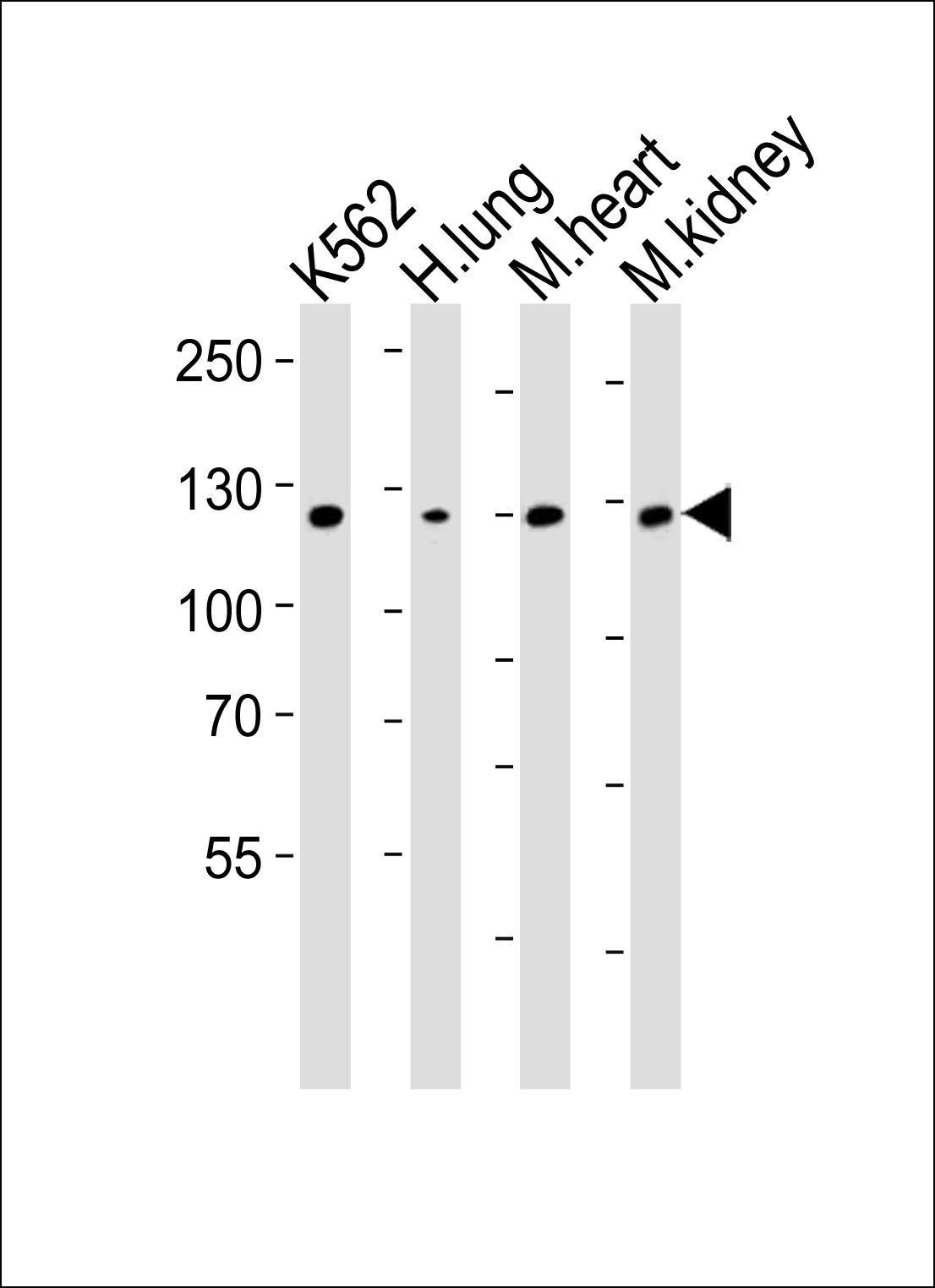
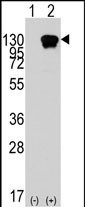
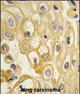
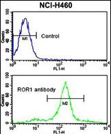
ROR1 Antibody
Purified Rabbit Polyclonal Antibody (Pab)
- 产品详情
- 文献引用 : 1
- 实验流程
- 背景知识
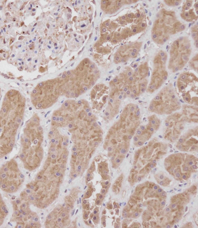
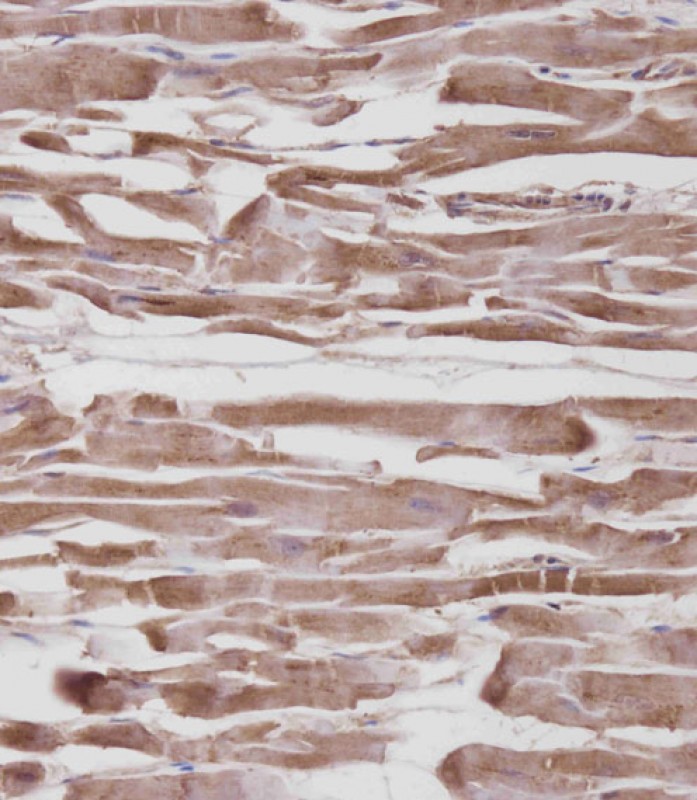
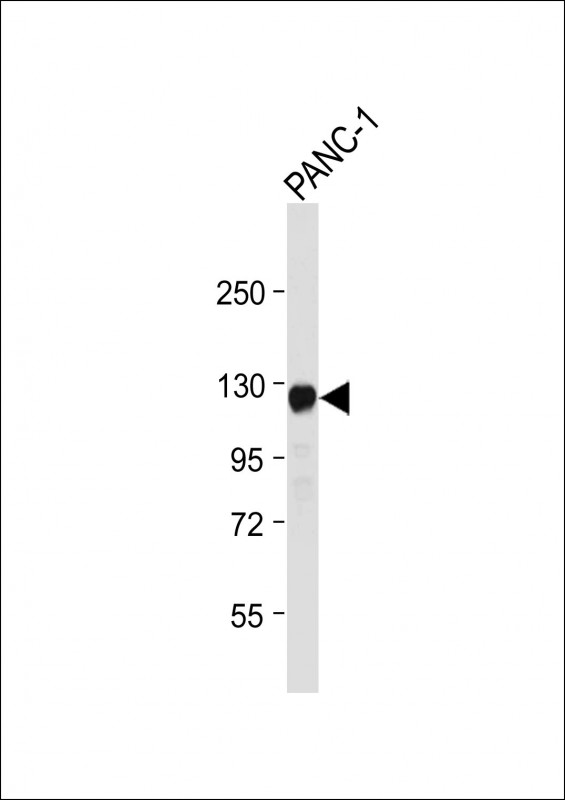
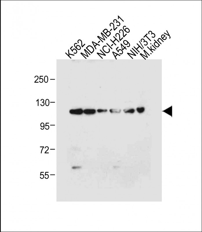
Application
| WB, IHC-P, FC, E |
|---|---|
| Primary Accession | Q01973 |
| Reactivity | Human, Mouse, Rat |
| Host | Rabbit |
| Clonality | Polyclonal |
| Isotype | Rabbit IgG |
| Calculated MW | 104283 Da |
| Gene ID | 4919 |
|---|---|
| Other Names | Tyrosine-protein kinase transmembrane receptor ROR1, Neurotrophic tyrosine kinase, receptor-related 1, ROR1, NTRKR1 |
| Target/Specificity | This ROR1 antibody is generated from rabbits immunized with recombinant human ROR1 protein (aa region: 112 - 399). |
| Dilution | WB~~1:1000 IHC-P~~1:100~500 FC~~1:10~50 E~~Use at an assay dependent concentration. |
| Format | Purified polyclonal antibody supplied in PBS with 0.09% (W/V) sodium azide. This antibody is purified through a protein A column, followed by peptide affinity purification. |
| Storage | Maintain refrigerated at 2-8°C for up to 2 weeks. For long term storage store at -20°C in small aliquots to prevent freeze-thaw cycles. |
| Precautions | ROR1 Antibody is for research use only and not for use in diagnostic or therapeutic procedures. |
| Name | ROR1 |
|---|---|
| Synonyms | NTRKR1 |
| Function | Has very low kinase activity in vitro and is unlikely to function as a tyrosine kinase in vivo (PubMed:25029443). Receptor for ligand WNT5A which activate downstream NFkB signaling pathway and may result in the inhibition of WNT3A-mediated signaling (PubMed:25029443, PubMed:27162350). In inner ear, crucial for spiral ganglion neurons to innervate auditory hair cells (PubMed:27162350). Via IGFBP5 ligand, forms a complex with ERBB2 to enhance CREB oncogenic signaling (PubMed:36949068). |
| Cellular Location | Membrane; Single- pass type I membrane protein. Cell projection, axon {ECO:0000250|UniProtKB:Q9Z139} |
| Tissue Location | Expressed strongly in human heart, lung and kidney, but weakly in the CNS. Isoform Short is strongly expressed in fetal and adult CNS and in a variety of human cancers, including those originating from CNS or PNS neuroectoderm |
For Research Use Only. Not For Use In Diagnostic Procedures.

Provided below are standard protocols that you may find useful for product applications.
BACKGROUND
ROR1 is a receptor protein tyrosine kinase whose cellular role has not been determined. It is a type I membrane protein and belongs to the ROR subfamily of cell surface receptors. Studies of a similar protein in mouse suggest that this protein may interact with another receptor protein tyrosine kinase and may be involved in skeletal and cardiac development.
REFERENCES
Nomi, M., et al., Mol. Cell. Biol. 21(24):8329-8335 (2001).
Reddy, U.R., et al., Genomics 41(2):283-285 (1997).
Reddy, U.R., et al., Oncogene 13(7):1555-1559 (1996).
Masiakowski, P., et al., J. Biol. Chem. 267(36):26181-26190 (1992).
终于等到您。ABCEPTA(百远生物)抗体产品。
点击下方“我要评价 ”按钮提交您的反馈信息,您的反馈和评价是我们最宝贵的财富之一,
我们将在1-3个工作日内处理您的反馈信息。
如有疑问,联系:0512-88856768 tech-china@abcepta.com.


