


 癌症的基本特征包括细胞增殖、血管生成、迁移、凋亡逃避机制和细胞永生等。找到癌症发生过程中这些通路的关键标记物和对应的抗体用于检测至关重要。
 为您推荐一个泛素化位点预测神器——泛素化分析工具,可以为您的蛋白的泛素化位点作出预测和评分。
 细胞自噬受体图形绘图工具为你的蛋白的细胞受体结合位点作出预测和评分,识别结合到自噬通路中的蛋白是非常重要的,便于让我们理解自噬在正常生理、病理过程中的作用,如发育、细胞分化、神经退化性疾病、压力条件下、感染和癌症。
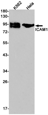
ICAM1 Rabbit mAb
- 产品详情
- 实验流程
- 背景知识
Application
| WB, IHC-P, FC, IP |
|---|---|
| Primary Accession | P05362 |
| Reactivity | Human |
| Host | Rabbit |
| Clonality | Monoclonal Antibody |
| Isotype | IgG |
| Conjugate | Unconjugated |
| Purification | Affinity Purified |
| Calculated MW | 57825 Da |
| Gene ID | 3383 |
|---|---|
| Other Names | ICAM1 |
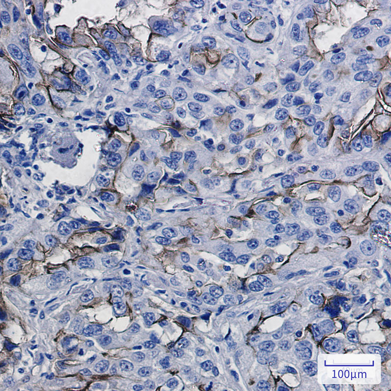
| Dilution | WB~~1:1000-1:5000 IHC-P~~1:50~200 FC~~1:50-1:100 IP~~1:10-1:100 |
| Format | Liquid in 50mM Tris-Glycine(pH 7.4), 0.15M NaCl, 40%Glycerol, 0.01% sodium azide and 0.05% BSA. |
| Storage | Store at 4°C short term. Aliquot and store at -20°C long term. Avoid freeze/thaw cycles. |
| Name | ICAM1 (HGNC:5344) |
|---|---|
| Function | Cell adhesion molecule that functions as a receptor ligand of the signaling receptor ITGAL:ITGB2/LFA-1 (lymphocyte-function associated (LFA) molecule 1) ensuring leukocyte cell-cell adhesion, by providing a calibrated system to namely adjust T-cell killing to the antigen stimulation strength (PubMed:3086451, PubMed:3340213, PubMed:38195629). Also functions as a ligand receptor of the signaling receptor ITGAM:ITGB2/MAC-1 ensuring adhesion between stimulated neutrophils and stimulated endothelial cells (PubMed:1980124). During leukocyte trans-endothelial migration, ICAM1 engagement promotes the assembly of endothelial apical cups through ARHGEF26/SGEF and RHOG activation (PubMed:17875742). Promotes cell aggregation in epithelial cells through interaction with MUC1 (PubMed:11173916). |
| Cellular Location | Cell membrane; Single-pass type I membrane protein |
| Tissue Location | Expressed on non-hematopoietic cells such as vascular endothelial cells, thymic epithelial cells, certain other epithelial cells, and fibroblasts, and on hematopoietic cells such as tissue macrophages, mitogen-stimulated T lymphocyte blasts, and germinal center dendritic cells in tonsils, lymph nodes, and Peyer's patches (PubMed:3086451). Expressed in low amounts on peripheral blood leukocytes (PubMed:3086451). |
For Research Use Only. Not For Use In Diagnostic Procedures.
Provided below are standard protocols that you may find useful for product applications.
BACKGROUND
CD54 is a 85-110 kD type I transmembrane protein also known as ICAM-1. It is expressed on activated endothelial cells, high endothelial venules, T and B cells, monocytes/macrophages, granulocytes, and dendritic cells. The expression of ICAM-1 can be released from the cell surface. CD54 plays a role in cellular adhesion and is involved in inflammation and leukocyte extravasation. CD54 has also been shown to be the major cellular receptor for rhinovirus. ICAM-1 binds to CD11a/CD18 (LFA-1), CD11b/CD18 (Mac-1), CD11c/CD18 (p150, 95) as well as hyaluronan and fibrinogen.
终于等到您。ABCEPTA(百远生物)抗体产品。
点击下方“我要评价 ”按钮提交您的反馈信息,您的反馈和评价是我们最宝贵的财富之一,
我们将在1-3个工作日内处理您的反馈信息。
如有疑问,联系:0512-88856768 tech-china@abcepta.com.


