


 癌症的基本特征包括细胞增殖、血管生成、迁移、凋亡逃避机制和细胞永生等。找到癌症发生过程中这些通路的关键标记物和对应的抗体用于检测至关重要。
 为您推荐一个泛素化位点预测神器——泛素化分析工具,可以为您的蛋白的泛素化位点作出预测和评分。
 细胞自噬受体图形绘图工具为你的蛋白的细胞受体结合位点作出预测和评分,识别结合到自噬通路中的蛋白是非常重要的,便于让我们理解自噬在正常生理、病理过程中的作用,如发育、细胞分化、神经退化性疾病、压力条件下、感染和癌症。
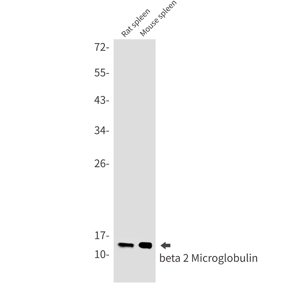
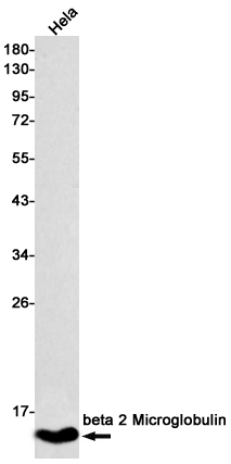
beta 2 Microglobulin Rabbit mAb
- 产品详情
- 实验流程
- 背景知识
Application
| WB, FC, IP |
|---|---|
| Primary Accession | P61769 |
| Reactivity | Rat, Human, Mouse |
| Host | Rabbit |
| Clonality | Monoclonal Antibody |
| Isotype | IgG |
| Conjugate | Unconjugated |
| Purification | Affinity Purified |
| Calculated MW | 13715 Da |
| Gene ID | 567 |
|---|---|
| Other Names | B2M |
| Dilution | WB~~1/500-1/1000 FC~~1:10~50 IP~~1/20 |
| Format | Liquid in 50mM Tris-Glycine(pH 7.4), 0.15M NaCl, 40%Glycerol, 0.01% sodium azide and 0.05% BSA. |
| Storage | Store at 4°C short term. Aliquot and store at -20°C long term. Avoid freeze/thaw cycles. |
| Name | B2M (HGNC:914) |
|---|---|
| Function | Component of the class I major histocompatibility complex (MHC). Involved in the presentation of peptide antigens to the immune system. Exogenously applied M.tuberculosis EsxA or EsxA-EsxB (or EsxA expressed in host) binds B2M and decreases its export to the cell surface (total protein levels do not change), probably leading to defects in class I antigen presentation (PubMed:25356553). |
| Cellular Location | Secreted. Cell surface. Note=Detected in serum and urine (PubMed:1336137, PubMed:7554280). {ECO:0000269|PubMed:7554280, ECO:0000269|Ref.6} |
For Research Use Only. Not For Use In Diagnostic Procedures.
Provided below are standard protocols that you may find useful for product applications.
BACKGROUND
Major histocompatibility complex (MHC) class 1 molecules bind to antigens for presentation on the surface of cells. The proteasome is responsible for producing these antigens from the components of foreign pathogens. MHC class 1 molecules consist of an a heavy chain that contains three subdomains (α1, α2, α3), and a non-covalent associating light chain, known as β-2-Microglobulin.
终于等到您。ABCEPTA(百远生物)抗体产品。
点击下方“我要评价 ”按钮提交您的反馈信息,您的反馈和评价是我们最宝贵的财富之一,
我们将在1-3个工作日内处理您的反馈信息。
如有疑问,联系:0512-88856768 tech-china@abcepta.com.


