


 癌症的基本特征包括细胞增殖、血管生成、迁移、凋亡逃避机制和细胞永生等。找到癌症发生过程中这些通路的关键标记物和对应的抗体用于检测至关重要。
 为您推荐一个泛素化位点预测神器——泛素化分析工具,可以为您的蛋白的泛素化位点作出预测和评分。
 细胞自噬受体图形绘图工具为你的蛋白的细胞受体结合位点作出预测和评分,识别结合到自噬通路中的蛋白是非常重要的,便于让我们理解自噬在正常生理、病理过程中的作用,如发育、细胞分化、神经退化性疾病、压力条件下、感染和癌症。
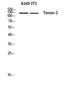
Tensin-2 Polyclonal Antibody
- 产品详情
- 实验流程
- 背景知识
Application
| WB |
|---|---|
| Primary Accession | Q63HR2 |
| Reactivity | Human, Mouse |
| Host | Rabbit |
| Clonality | Polyclonal |
| Calculated MW | 152580 Da |
| Gene ID | 23371 |
|---|---|
| Other Names | TENC1; KIAA1075; TNS2; Tensin-like C1 domain-containing phosphatase; C1 domain-containing phosphatase and tensin homolog; C1-TEN; Tensin-2 |
| Dilution | WB~~Western Blot: 1/500 - 1/2000. ELISA: 1/10000. Not yet tested in other applications. |
| Format | Liquid in PBS containing 50% glycerol, 0.5% BSA and 0.09% (W/V) sodium azide. |
| Storage Conditions | -20℃ |
| Name | TNS2 |
|---|---|
| Synonyms | KIAA1075, TENC1 |
| Function | Tyrosine-protein phosphatase which regulates cell motility, proliferation and muscle-response to insulin (PubMed:15817639, PubMed:23401856). Phosphatase activity is mediated by binding to phosphatidylinositol-3,4,5-triphosphate (PtdIns(3,4,5)P3) via the SH2 domain (PubMed:30092354). In muscles and under catabolic conditions, dephosphorylates IRS1 leading to its degradation and muscle atrophy (PubMed:23401856, PubMed:30092354). Negatively regulates PI3K-AKT pathway activation (PubMed:15817639, PubMed:23401856, PubMed:30092354). Dephosphorylates nephrin NPHS1 in podocytes which regulates activity of the mTORC1 complex (PubMed:28955049). Under normal glucose conditions, NPHS1 outcompetes IRS1 for binding to phosphatidylinositol 3-kinase (PI3K) which balances mTORC1 activity but high glucose conditions lead to up-regulation of TNS2, increased NPHS1 dephosphorylation and activation of mTORC1, contributing to podocyte hypertrophy and proteinuria (PubMed:28955049). Required for correct podocyte morphology, podocyte-glomerular basement membrane interaction and integrity of the glomerular filtration barrier (By similarity). Enhances RHOA activation in the presence of DLC1 (PubMed:26427649). Plays a role in promoting DLC1-dependent remodeling of the extracellular matrix (PubMed:20069572). |
| Cellular Location | Cell junction, focal adhesion. Cell membrane; Peripheral membrane protein; Cytoplasmic side. Cytoplasm. Note=Detected at the end of actin stress fibers. Detected in cytoplasmic punctate bodies (PubMed:22019427, PubMed:25101860). Localizes to both focal adhesions and fibrillar adhesions but is found mainly in focal adhesions (PubMed:20069572) Enriched in dynamic focal adhesions at the leading edge of the cell and is found only rarely in fibrillar adhesions on the ventral surface of cells (PubMed:20069572). |
| Tissue Location | Detected in heart, kidney, brain, thymus, spleen, liver, placenta, lung, skeletal muscle and small intestine |
Research Areas
For Research Use Only. Not For Use In Diagnostic Procedures.
Application Protocols
Provided below are standard protocols that you may find useful for product applications.
BACKGROUND
Regulates cell motility and proliferation. May have phosphatase activity. Reduces AKT1 phosphorylation. Lowers AKT1 kinase activity and interferes with AKT1 signaling.
终于等到您。ABCEPTA(百远生物)抗体产品。
点击下方“我要评价 ”按钮提交您的反馈信息,您的反馈和评价是我们最宝贵的财富之一,
我们将在1-3个工作日内处理您的反馈信息。
如有疑问,联系:0512-88856768 tech-china@abcepta.com.
¥ 1,500.00
Cat# AP73842


