


 癌症的基本特征包括细胞增殖、血管生成、迁移、凋亡逃避机制和细胞永生等。找到癌症发生过程中这些通路的关键标记物和对应的抗体用于检测至关重要。
 为您推荐一个泛素化位点预测神器——泛素化分析工具,可以为您的蛋白的泛素化位点作出预测和评分。
 细胞自噬受体图形绘图工具为你的蛋白的细胞受体结合位点作出预测和评分,识别结合到自噬通路中的蛋白是非常重要的,便于让我们理解自噬在正常生理、病理过程中的作用,如发育、细胞分化、神经退化性疾病、压力条件下、感染和癌症。
PKC nu Antibody
Purified Rabbit Polyclonal Antibody (Pab)

- 产品详情
- 文献引用 : 1
- 实验流程
- 背景知识
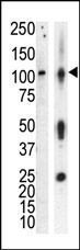
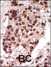
Application
| WB, IHC-P, E |
|---|---|
| Primary Accession | O94806 |
| Reactivity | Mouse, Human |
| Host | Rabbit |
| Clonality | Polyclonal |
| Isotype | Rabbit IgG |
| Calculated MW | 100471 Da |
| Antigen Region | 352-384 aa |
| Gene ID | 23683 |
|---|---|
| Other Names | Serine/threonine-protein kinase D3, Protein kinase C nu type, Protein kinase EPK2, nPKC-nu, PRKD3, EPK2, PRKCN |
| Target/Specificity | This PKC-nu antibody is generated from rabbits immunized with a KLH conjugated synthetic peptide between 352-384 amino acids from human PKC-nu. |
| Dilution | WB~~1:1000 IHC-P~~1:100~500 E~~Use at an assay dependent concentration. |
| Format | Purified polyclonal antibody supplied in PBS with 0.09% (W/V) sodium azide. This antibody is prepared by Saturated Ammonium Sulfate (SAS) precipitation followed by dialysis against PBS. |
| Storage | Maintain refrigerated at 2-8°C for up to 2 weeks. For long term storage store at -20°C in small aliquots to prevent freeze-thaw cycles. |
| Precautions | PKC nu Antibody is for research use only and not for use in diagnostic or therapeutic procedures. |
| Name | PRKD3 |
|---|---|
| Synonyms | EPK2, PRKCN |
| Function | Converts transient diacylglycerol (DAG) signals into prolonged physiological effects, downstream of PKC. Involved in resistance to oxidative stress (By similarity). |
| Cellular Location | Cytoplasm. Membrane. Note=Translocation to the cell membrane is required for kinase activation |
| Tissue Location | Ubiquitous. |
For Research Use Only. Not For Use In Diagnostic Procedures.

Provided below are standard protocols that you may find useful for product applications.
BACKGROUND
Protein kinase C (PKC) is a family of serine- and threonine-specific protein kinases that can be activated by calcium and second messenger diacylglycerol. PKC family members phosphorylate a wide variety of protein targets and are known to be involved in diverse cellular signaling pathways. PKC also serve as major receptors for phorbol esters, a class of tumor promoters. Each member of the PKC family has a specific expression profile and is believed to play distinct roles in cells. PKC nu is one of the PKC family members. This kinase can be activated rapidly by the agonists of G protein-coupled receptors. It resides in both cytoplasm and nucleus, and its nuclear accumulation is found to be dramatically enhanced in response to its activation. This kinase can also be activated after B-cell antigen receptor (BCR) engagement, which requires intact phopholipase C gamma and the involvement of other PKC family members.
REFERENCES
Yeaman, C., et al., Nat. Cell Biol. 6(2):106-112 (2004).
Rey, O., et al., J. Biol. Chem. 278(26):23773-23785 (2003).
Matthews, S.A., et al., J. Biol. Chem. 278(11):9086-9091 (2003).
Bennasser, Y., et al., Virology 303(1):174-180 (2002).
Bennasser, Y., et al., FASEB J. 16(6):546-554 (2002).
终于等到您。ABCEPTA(百远生物)抗体产品。
点击下方“我要评价 ”按钮提交您的反馈信息,您的反馈和评价是我们最宝贵的财富之一,
我们将在1-3个工作日内处理您的反馈信息。
如有疑问,联系:0512-88856768 tech-china@abcepta.com.


