


 癌症的基本特征包括细胞增殖、血管生成、迁移、凋亡逃避机制和细胞永生等。找到癌症发生过程中这些通路的关键标记物和对应的抗体用于检测至关重要。
 为您推荐一个泛素化位点预测神器——泛素化分析工具,可以为您的蛋白的泛素化位点作出预测和评分。
 细胞自噬受体图形绘图工具为你的蛋白的细胞受体结合位点作出预测和评分,识别结合到自噬通路中的蛋白是非常重要的,便于让我们理解自噬在正常生理、病理过程中的作用,如发育、细胞分化、神经退化性疾病、压力条件下、感染和癌症。
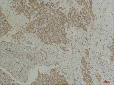
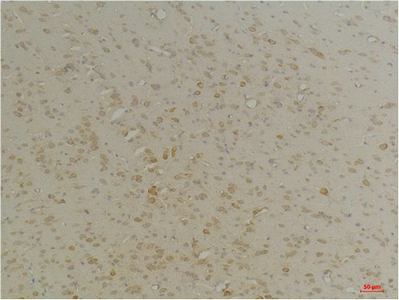
CLIC4 Polyclonal Antibody

- 产品详情
- 实验流程
- 背景知识
Application
| WB, IHC-P |
|---|---|
| Primary Accession | Q9Y696 |
| Reactivity | Human, Rat, Mouse |
| Host | Rabbit |
| Clonality | Polyclonal |
| Calculated MW | 28772 Da |
| Gene ID | 25932 |
|---|---|
| Other Names | CLIC4; Chloride intracellular channel protein 4; Intracellular chloride ion channel protein p64H1 |
| Dilution | WB~~WB 1:1000-2000, IHC 1:100-200 IHC-P~~WB 1:1000-2000, IHC 1:100-200 |
| Format | Liquid in PBS containing 50% glycerol, 0.5% BSA and 0.09% (W/V) sodium azide. |
| Storage Conditions | -20℃ |
| Name | CLIC4 {ECO:0000303|PubMed:12163372, ECO:0000312|HGNC:HGNC:13518} |
|---|---|
| Function | In the soluble state, catalyzes glutaredoxin-like thiol disulfide exchange reactions with reduced glutathione as electron donor (PubMed:25581026, PubMed:37759794). Can insert into membranes and form voltage-dependent multi-ion conductive channels. Membrane insertion seems to be redox-regulated and may occur only under oxidizing conditions (By similarity) (PubMed:16176272). Has alternate cellular functions like a potential role in angiogenesis or in maintaining apical-basolateral membrane polarity during mitosis and cytokinesis. Could also promote endothelial cell proliferation and regulate endothelial morphogenesis (tubulogenesis). Promotes cell-surface expression of HRH3. |
| Cellular Location | Cytoplasm, cytoskeleton, microtubule organizing center, centrosome. Cytoplasmic vesicle membrane; Single-pass membrane protein. Nucleus. Cell membrane; Single-pass membrane protein. Mitochondrion {ECO:0000250|UniProtKB:Q9Z0W7}. Cell junction. Endoplasmic reticulum membrane {ECO:0000250|UniProtKB:Q9Z0W7}; Single-pass membrane protein {ECO:0000250|UniProtKB:Q9Z0W7}. Note=Colocalized with AKAP9 at the centrosome and midbody. Exists both as soluble cytoplasmic protein and as membrane protein with probably a single transmembrane domain Present in an intracellular vesicular compartment that likely represent trans-Golgi network vesicles. Might not be present in the nucleus of cardiac cells. {ECO:0000250|UniProtKB:Q9Z0W7, ECO:0000269|PubMed:14569596} |
| Tissue Location | Detected in epithelial cells from colon, esophagus and kidney (at protein level). Expression is prominent in heart, kidney, placenta and skeletal muscle. |
For Research Use Only. Not For Use In Diagnostic Procedures.
Provided below are standard protocols that you may find useful for product applications.
BACKGROUND
Can insert into membranes and form poorly selective ion channels that may also transport chloride ions. Channel activity depends on the pH. Membrane insertion seems to be redox-regulated and may occur only under oxydizing conditions. Promotes cell- surface expression of HRH3. Has alternate cellular functions like a potential role in angiogenesis or in maintaining apical- basolateral membrane polarity during mitosis and cytokinesis. Could also promote endothelial cell proliferation and regulate endothelial morphogenesis (tubulogenesis).
终于等到您。ABCEPTA(百远生物)抗体产品。
点击下方“我要评价 ”按钮提交您的反馈信息,您的反馈和评价是我们最宝贵的财富之一,
我们将在1-3个工作日内处理您的反馈信息。
如有疑问,联系:0512-88856768 tech-china@abcepta.com.


