Myosin VIIa Polyclonal Antibody
Purified Rabbit Polyclonal Antibody (Pab)
- 产品详情
- 实验流程
Application
| IHC-P, IHC-F, IF, E |
|---|---|
| Primary Accession | Q13402 |
| Reactivity | Rat, Pig, Dog, Bovine |
| Host | Rabbit |
| Clonality | Polyclonal |
| Calculated MW | 254390 Da |
| Physical State | Liquid |
| Immunogen | KLH conjugated synthetic peptide derived from human Myosin VIIa |
| Epitope Specificity | 851-950/2215 |
| Isotype | IgG |
| Purity | affinity purified by Protein A |
| Buffer | 0.01M TBS (pH7.4) with 1% BSA, 0.02% Proclin300 and 50% Glycerol. |
| SUBCELLULAR LOCATION | Cytoplasm (Probable). Note=In the photoreceptor cells, mainly localized in the inner and base of outer segments as well as in the synaptic ending region. |
| SIMILARITY | Contains 2 FERM domains. Contains 5 IQ domains. Contains 1 myosin head-like domain. Contains 2 MyTH4 domains. Contains 1 SH3 domain. |
| SUBUNIT | Interacts with PLEKHB1 (via PH domain). Might homodimerize in a two headed molecule through the formation of a coiled-coil rod. Binds MYRIP and WHRN. |
| DISEASE | Defects in MYO7A are the cause of Usher syndrome type 1B (USH1B) [MIM:276900]. USH is a genetically heterogeneous condition characterized by the association of retinitis pigmentosa and sensorineural deafness. Age at onset and differences in auditory and vestibular function distinguish Usher syndrome type 1 (USH1), Usher syndrome type 2 (USH2) and Usher syndrome type 3 (USH3). USH1 is characterized by profound congenital sensorineural deafness, absent vestibular function and prepubertal onset of progressive retinitis pigmentosa leading to blindness. Defects in MYO7A are the cause of deafness autosomal recessive type 2 (DFNB2) [MIM:600060]; also called neurosensory non-syndromic recessive deafness 2 (NSRD2). DFNB2 is a form of sensorineural hearing loss. Sensorineural deafness results from damage to the neural receptors of the inner ear, the nerve pathways to the brain, or the area of the brain that receives sound information. Defects in MYO7A are the cause of deafness autosomal dominant type 11 (DFNA11) [MIM:601317]. |
| Important Note | This product as supplied is intended for research use only, not for use in human, therapeutic or diagnostic applications. |
| Background Descriptions | Myosins are actin-based motor molecules with ATPase activity. Unconventional myosins serve in intracellular movements. Their highly divergent tails are presumed to bind to membranous compartments, which would be moved relative to actin filaments. In retina, myosin VIIa may play a role in trafficking of ribbon-synaptic vesicle complexes and renewal of the outer photoreceptors disks. In inner ear, it may maintain the rigidity of stereocilia during the dynamic movements of the bundle. |
| Gene ID | 4647 |
|---|---|
| Other Names | Unconventional myosin-VIIa, MYO7A, USH1B |
| Target/Specificity | Expressed in the pigment epithelium and the photoreceptor cells of the retina. Also found in kidney, liver, testis, cochlea, lymphocytes. Not expressed in brain. |
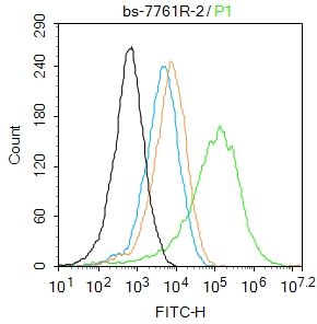
| Dilution | IHC-P=1:100-500,IHC-F=1:100-500,IF=1:100-500,Flow-Cyt=2ug/Test,ELISA=1:5000-10000 |
| Format | 0.01M TBS(pH7.4) with 1% BSA, 0.09% (W/V) sodium azide and 50% Glyce |
| Storage | Store at -20 °C for one year. Avoid repeated freeze/thaw cycles. When reconstituted in sterile pH 7.4 0.01M PBS or diluent of antibody the antibody is stable for at least two weeks at 2-4 °C. |
| Name | MYO7A (HGNC:7606) |
|---|---|
| Synonyms | USH1B |
| Function | Myosins are actin-based motor molecules with ATPase activity. Unconventional myosins serve in intracellular movements. Their highly divergent tails bind to membranous compartments, which are then moved relative to actin filaments. In the retina, plays an important role in the renewal of the outer photoreceptor disks. Plays an important role in the distribution and migration of retinal pigment epithelial (RPE) melanosomes and phagosomes, and in the regulation of opsin transport in retinal photoreceptors. In the inner ear, plays an important role in differentiation, morphogenesis and organization of cochlear hair cell bundles. Involved in hair-cell vesicle trafficking of aminoglycosides, which are known to induce ototoxicity (By similarity). Motor protein that is a part of the functional network formed by USH1C, USH1G, CDH23 and MYO7A that mediates mechanotransduction in cochlear hair cells. Required for normal hearing. |
| Cellular Location | Cytoplasm {ECO:0000250|UniProtKB:P97479}. Cytoplasm, cell cortex {ECO:0000250|UniProtKB:P97479}. Cytoplasm, cytoskeleton {ECO:0000250|UniProtKB:P97479}. Synapse. Note=In the photoreceptor cells, mainly localized in the inner and base of outer segments as well as in the synaptic ending region (PubMed:8842737). In retinal pigment epithelial cells colocalizes with a subset of melanosomes, displays predominant localization to stress fiber-like structures and some localization to cytoplasmic puncta (PubMed:19643958, PubMed:27331610). Detected at the tip of cochlear hair cell stereocilia (PubMed:21709241). The complex formed by MYO7A, USH1C and USH1G colocalizes with F-actin (PubMed:21709241). |
| Tissue Location | Expressed in the pigment epithelium and the photoreceptor cells of the retina. Also found in kidney, liver, testis, cochlea, lymphocytes. Not expressed in brain |
Research Areas
For Research Use Only. Not For Use In Diagnostic Procedures.
Application Protocols
Provided below are standard protocols that you may find useful for product applications.
终于等到您。ABCEPTA(百远生物)抗体产品。
点击下方“我要评价 ”按钮提交您的反馈信息,您的反馈和评价是我们最宝贵的财富之一,
我们将在1-3个工作日内处理您的反馈信息。
如有疑问,联系:0512-88856768 tech-china@abcepta.com.
¥ 1,500.00
Cat# AP58748


 癌症的基本特征包括细胞增殖、血管生成、迁移、凋亡逃避机制和细胞永生等。找到癌症发生过程中这些通路的关键标记物和对应的抗体用于检测至关重要。
 为您推荐一个泛素化位点预测神器——泛素化分析工具,可以为您的蛋白的泛素化位点作出预测和评分。
 细胞自噬受体图形绘图工具为你的蛋白的细胞受体结合位点作出预测和评分,识别结合到自噬通路中的蛋白是非常重要的,便于让我们理解自噬在正常生理、病理过程中的作用,如发育、细胞分化、神经退化性疾病、压力条件下、感染和癌症。


