


 癌症的基本特征包括细胞增殖、血管生成、迁移、凋亡逃避机制和细胞永生等。找到癌症发生过程中这些通路的关键标记物和对应的抗体用于检测至关重要。
 为您推荐一个泛素化位点预测神器——泛素化分析工具,可以为您的蛋白的泛素化位点作出预测和评分。
 细胞自噬受体图形绘图工具为你的蛋白的细胞受体结合位点作出预测和评分,识别结合到自噬通路中的蛋白是非常重要的,便于让我们理解自噬在正常生理、病理过程中的作用,如发育、细胞分化、神经退化性疾病、压力条件下、感染和癌症。
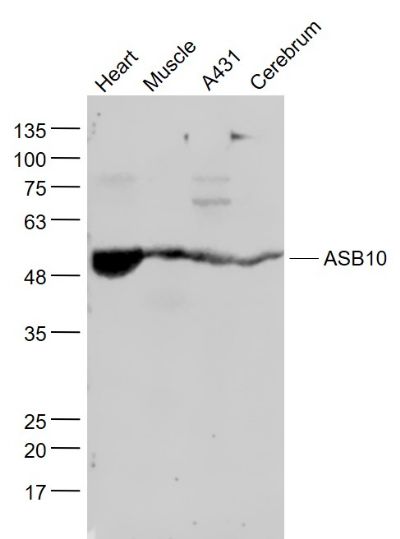
ASB10 Polyclonal Antibody
Purified Rabbit Polyclonal Antibody (Pab)
- 产品详情
- 实验流程
Application
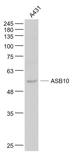
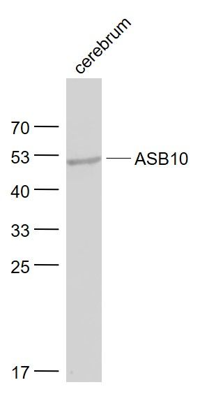
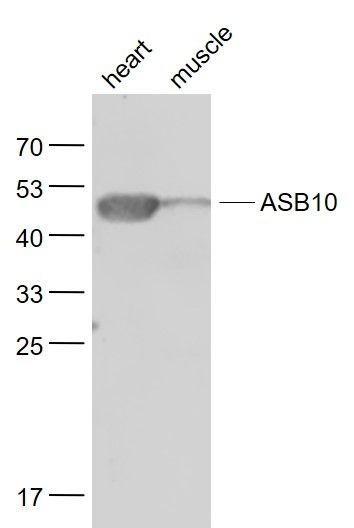
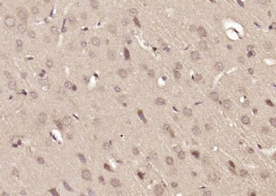
| WB, IHC-P, IHC-F, IF, E |
|---|---|
| Primary Accession | Q8WXI3 |
| Reactivity | Rat, Pig, Dog, Bovine |
| Host | Rabbit |
| Clonality | Polyclonal |
| Calculated MW | 50894 Da |
| Physical State | Liquid |
| Immunogen | KLH conjugated synthetic peptide derived from human ASB10 |
| Epitope Specificity | 401-450/467 |
| Isotype | IgG |
| Purity | affinity purified by Protein A |
| Buffer | 0.01M TBS (pH7.4) with 1% BSA, 0.02% Proclin300 and 50% Glycerol. |
| SIMILARITY | Contains 7 ANK repeats.Contains 1 SOCS box domain. |
| Important Note | This product as supplied is intended for research use only, not for use in human, therapeutic or diagnostic applications. |
| Background Descriptions | ASB10 is a member of the ankyrin repeat and SOCS box-containing (ASB) family of proteins. SOCS boxes are carboxy terminal regions of homology found in the suppressor of cytokine signaling (SOCS) family of proteins. The box region is thought to be the point of interaction between SOCS proteins and E3 ubiquitin ligases. The SOCS box couples the suppressor of cytokine signalling proteins and their binding partners with the elongin B and C complex, possibly targeting them for degradation. |
| Gene ID | 136371 |
|---|---|
| Other Names | Ankyrin repeat and SOCS box protein 10, ASB-10, ASB10 |
| Dilution | WB=1:500-2000,IHC-P=1:100-500,IHC-F=1:100-500,IF=1:100-500,ELISA=1:5000-10000 |
| Format | 0.01M TBS(pH7.4) with 1% BSA, 0.09% (W/V) sodium azide and 50% Glyce |
| Storage | Store at -20 °C for one year. Avoid repeated freeze/thaw cycles. When reconstituted in sterile pH 7.4 0.01M PBS or diluent of antibody the antibody is stable for at least two weeks at 2-4 °C. |
| Name | ASB10 |
|---|---|
| Function | May be a substrate-recognition component of a SCF-like ECS (Elongin-Cullin-SOCS-box protein) E3 ubiquitin-protein ligase complex which mediates the ubiquitination and subsequent proteasomal degradation of target proteins. |
| Cellular Location | Cytoplasm. Nucleus. Note=In the ciliary body, it is detected in the cytoplasm and perinuclear region of the pigmented ciliary epithelial layer. In the retina, it is detected in the nuclei of retinal ganglion cells |
| Tissue Location | Expressed in the eye. The highest expression is observed in the iris, with moderate levels in the trabecular meshwork (TM), the lamina, and the optic nerve; slightly lower levels in the ciliary body, retina, and choroid; and very low levels in the lens |
Research Areas
For Research Use Only. Not For Use In Diagnostic Procedures.
Application Protocols
Provided below are standard protocols that you may find useful for product applications.
终于等到您。ABCEPTA(百远生物)抗体产品。
点击下方“我要评价 ”按钮提交您的反馈信息,您的反馈和评价是我们最宝贵的财富之一,
我们将在1-3个工作日内处理您的反馈信息。
如有疑问,联系:0512-88856768 tech-china@abcepta.com.


