


 癌症的基本特征包括细胞增殖、血管生成、迁移、凋亡逃避机制和细胞永生等。找到癌症发生过程中这些通路的关键标记物和对应的抗体用于检测至关重要。
 为您推荐一个泛素化位点预测神器——泛素化分析工具,可以为您的蛋白的泛素化位点作出预测和评分。
 细胞自噬受体图形绘图工具为你的蛋白的细胞受体结合位点作出预测和评分,识别结合到自噬通路中的蛋白是非常重要的,便于让我们理解自噬在正常生理、病理过程中的作用,如发育、细胞分化、神经退化性疾病、压力条件下、感染和癌症。
FUT2 Rabbit pAb
FUT2 Rabbit pAb
- 产品详情
- 实验流程
- 背景知识
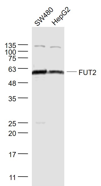
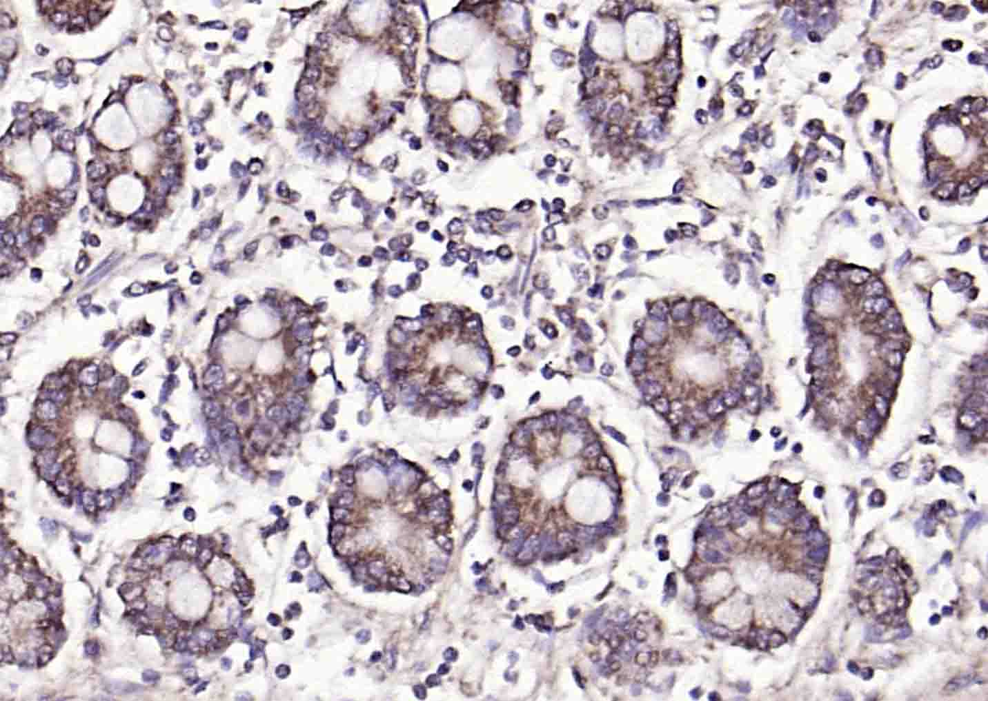
Application
| WB, IHC-P, IHC-F, IF |
|---|---|
| Primary Accession | Q10981 |
| Reactivity | Human |
| Predicted | Mouse, Rat, Pig, Horse, Rabbit, Sheep |
| Host | Rabbit |
| Clonality | Polyclonal |
| Calculated MW | 39017 Da |
| Physical State | Liquid |
| Immunogen | KLH conjugated synthetic peptide derived from Human FUT2 |
| Epitope Specificity | 231-343/343 |
| Isotype | IgG |
| Purity | affinity purified by Protein A |
| Buffer | 0.01M TBS (pH7.4) with 1% BSA, 0.02% Proclin300 and 50% Glycerol. |
| SUBCELLULAR LOCATION | Golgi apparatus, Golgi stack membrane; Single-pass type II membrane protein. Note=Membrane-bound form in trans cisternae of Golgi. |
| SIMILARITY | Belongs to the glycosyltransferase 11 family. |
| Important Note | This product as supplied is intended for research use only, not for use in human, therapeutic or diagnostic applications. |
| Background Descriptions | Creates a soluble precursor oligosaccharide FuC-alpha ((1,2)Galbeta-) called the H antigen which is an essential substrate for the final step in the soluble A and B antigen synthesis pathway. H and Se enzymes fucosylate the same acceptor substrates but exhibit different Km values. FUT2 is expressed on the surface of several human tumor cell lines such as BEL-7404, SPC-A-1, and SGC-7901. |
| Gene ID | 2524 |
|---|---|
| Other Names | Galactoside alpha-(1, 2)-fucosyltransferase 2, Alpha(1, 2)FT 2, Fucosyltransferase 2, GDP-L-fucose:beta-D-galactoside 2-alpha-L-fucosyltransferase 2, SE2, Secretor blood group alpha-2-fucosyltransferase, Secretor factor, Se, Type 1 galactoside alpha-(1, 2)-fucosyltransferase FUT2, 2.4.1.69, Type 2 galactoside alpha-(1, 2)-fucosyltransferase FUT2, 2.4.1.344, FUT2 (HGNC:4013), SEC2 |
| Target/Specificity | Small intestine, colon and lung. |
| Dilution | WB=1:500-2000,IHC-P=1:100-500,IHC-F=1:100-500,IF=1:100-500 |
| Storage | Store at -20 °C for one year. Avoid repeated freeze/thaw cycles. When reconstituted in sterile pH 7.4 0.01M PBS or diluent of antibody the antibody is stable for at least two weeks at 2-4 °C. |
| Name | FUT2 (HGNC:4013) |
|---|---|
| Synonyms | SEC2 |
| Function | Catalyzes the transfer of L-fucose, from a guanosine diphosphate-beta-L-fucose, to the terminal galactose on both O- and N- linked glycans chains of cell surface glycoproteins and glycolipids and the resulting epitope regulates several processes such as cell-cell interaction including host-microbe interaction, cell surface expression and cell proliferation (PubMed:12692541, PubMed:7876235, PubMed:8018146). Preferentially fucosylates gangliosides GA1 and GM1 in the antrum, cecum and colon and in the female reproductive organs (By similarity). Fucosylated host glycoproteins or glycolipids mediate interaction with intestinal microbiota influencing its composition (PubMed:21625510, PubMed:22068912, PubMed:24733310). Creates a soluble precursor oligosaccharide FuC-alpha ((1,2)Galbeta-) called the H antigen which is an essential substrate for the final step in the soluble ABO blood group antigen synthesis pathway (PubMed:7876235). |
| Cellular Location | Golgi apparatus, Golgi stack membrane; Single- pass type II membrane protein. Note=Membrane-bound form in trans cisternae of Golgi |
| Tissue Location | Small intestine, colon and lung. |
For Research Use Only. Not For Use In Diagnostic Procedures.
Provided below are standard protocols that you may find useful for product applications.
BACKGROUND
Creates a soluble precursor oligosaccharide FuC-alpha ((1,2)Galbeta-) called the H antigen which is an essential substrate for the final step in the soluble A and B antigen synthesis pathway. H and Se enzymes fucosylate the same acceptor substrates but exhibit different Km values. FUT2 is expressed on the surface of several human tumor cell lines such as BEL-7404, SPC-A-1, and SGC-7901.
终于等到您。ABCEPTA(百远生物)抗体产品。
点击下方“我要评价 ”按钮提交您的反馈信息,您的反馈和评价是我们最宝贵的财富之一,
我们将在1-3个工作日内处理您的反馈信息。
如有疑问,联系:0512-88856768 tech-china@abcepta.com.


