


 癌症的基本特征包括细胞增殖、血管生成、迁移、凋亡逃避机制和细胞永生等。找到癌症发生过程中这些通路的关键标记物和对应的抗体用于检测至关重要。
 为您推荐一个泛素化位点预测神器——泛素化分析工具,可以为您的蛋白的泛素化位点作出预测和评分。
 细胞自噬受体图形绘图工具为你的蛋白的细胞受体结合位点作出预测和评分,识别结合到自噬通路中的蛋白是非常重要的,便于让我们理解自噬在正常生理、病理过程中的作用,如发育、细胞分化、神经退化性疾病、压力条件下、感染和癌症。
MAGI1 Polyclonal Antibody
Purified Rabbit Polyclonal Antibody (Pab)
- 产品详情
- 实验流程
Application
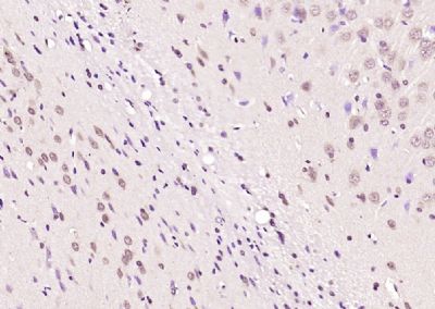
| WB, IHC-P, IHC-F, IF, ICC, E |
|---|---|
| Primary Accession | Q96QZ7 |
| Reactivity | Rat, Pig, Dog, Bovine |
| Host | Rabbit |
| Clonality | Polyclonal |
| Calculated MW | 164581 Da |
| Physical State | Liquid |
| Immunogen | KLH conjugated synthetic peptide derived from human MAGI1 |
| Epitope Specificity | 161-260/1491 |
| Isotype | IgG |
| Purity | affinity purified by Protein A |
| Buffer | 0.01M TBS (pH7.4) with 1% BSA, 0.02% Proclin300 and 50% Glycerol. |
| SUBCELLULAR LOCATION | Cell junction; cell membrane; tight junction; peripheral membrane protein. Localizes to epithelial cells tight junctions. |
| SIMILARITY | Contains 1 guanylate kinase-like domain. Contains 6 PDZ (DHR) domains. Contains 2 WW domains. |
| SUBUNIT | Interacts through its WW 2 domain with SYNPO and through its PDZ 5 domain with ACTN4. Interacts with cytoplasmic domain of BAI1. Interacts via its WW domains with DRPLA. Interacts with ESAM, LRP2 and CXADR. May interact with CTNNB1. Interacts through its PDZ 1 domain with NET1 (By similarity). Interacts with ASIC3 and AMOT. Interacts with FCHSD2. Interacts with IGSF5/JAM4 and through its PDZ 2 and 3 domains with NPHS1 forming a tripartite complex (By similarity). Interacts with DDN. |
| Important Note | This product as supplied is intended for research use only, not for use in human, therapeutic or diagnostic applications. |
| Background Descriptions | The membrane-associated guanylate kinase (MAGUK) proteins are concentrated at the membrane-cytoskeletal interface where they facilitate the assembly of multiprotein complexes on the inner surface of the plasma membrane. Three protein-protein interaction modules characteristically define MAGUK related proteins: the PDZ domain, the SH3 domain and the guanylate kinase (GuK) domain. The closely related MAGUK proteins, MAGI-1, MAGI-2 and MAGI-3 (membrane associated guanylate kinase inverted-1 and 2), likewise contain the GuK domain and five PDZ domains; however, the SH3 domain is replaced with a WW domain. The transcripts of MAGI-1 are alternatively spliced to produce three distinct proteins having unique C-terminals. Two variants, MAGI-1a and MAGI-1b, are associated with the membrane and cytosolic fractions and are primarily expressed in the brain. The third isoform, MAGI-1c, encodes for a nuclear localization signal that localizes MAGI-1c to the nucleus, and it is primarily expressed in the liver and kidney. MAGI-2 and MAGI-3 are localized to the plasma membrane, and they contribute to protein scaffolding by associating with the protein phosphatase PTEN. |
| Gene ID | 9223 |
|---|---|
| Other Names | Membrane-associated guanylate kinase, WW and PDZ domain-containing protein 1, Atrophin-1-interacting protein 3, AIP-3, BAI1-associated protein 1, BAP-1, Membrane-associated guanylate kinase inverted 1, MAGI-1, Trinucleotide repeat-containing gene 19 protein, WW domain-containing protein 3, WWP3, MAGI1, AIP3, BAIAP1, BAP1, TNRC19 |
| Target/Specificity | Widely expressed with the exception of skeletal muscle. Isoform 1, isoform 2 and isoform 6 are highly expressed in colon, kidney, lung, liver, and pancreas. Isoform 5 is predominantly expressed in brain and heart. Isoform 3 and isoform 4 are highly expressed in pancreas and brain. |
| Dilution | WB=1:500-2000,IHC-P=1:100-500,IHC-F=1:100-500,ICC=1:100-500,IF=1:100-500,ELISA=1:5000-10000 |
| Format | 0.01M TBS(pH7.4) with 1% BSA, 0.09% (W/V) sodium azide and 50% Glyce |
| Storage | Store at -20 °C for one year. Avoid repeated freeze/thaw cycles. When reconstituted in sterile pH 7.4 0.01M PBS or diluent of antibody the antibody is stable for at least two weeks at 2-4 °C. |
| Name | MAGI1 |
|---|---|
| Synonyms | AIP3, BAIAP1, BAP1, TNRC19 |
| Function | Plays a role in coupling actin fibers to cell junctions in endothelial cells, via its interaction with AMOTL2 and CDH5 (By similarity). May regulate acid-induced ASIC3 currents by modulating its expression at the cell surface (By similarity). |
| Cellular Location | Cell junction, tight junction. Cell membrane; Peripheral membrane protein. Note=Localizes to epithelial cells tight junctions |
| Tissue Location | Widely expressed with the exception of skeletal muscle. Isoform 1, isoform 2 and isoform 6 are highly expressed in colon, kidney, lung, liver, and pancreas. Isoform 5 is predominantly expressed in brain and heart. Isoform 3 and isoform 4 are highly expressed in pancreas and brain. |
Research Areas
For Research Use Only. Not For Use In Diagnostic Procedures.
Application Protocols
Provided below are standard protocols that you may find useful for product applications.
终于等到您。ABCEPTA(百远生物)抗体产品。
点击下方“我要评价 ”按钮提交您的反馈信息,您的反馈和评价是我们最宝贵的财富之一,
我们将在1-3个工作日内处理您的反馈信息。
如有疑问,联系:0512-88856768 tech-china@abcepta.com.


