


 癌症的基本特征包括细胞增殖、血管生成、迁移、凋亡逃避机制和细胞永生等。找到癌症发生过程中这些通路的关键标记物和对应的抗体用于检测至关重要。
 为您推荐一个泛素化位点预测神器——泛素化分析工具,可以为您的蛋白的泛素化位点作出预测和评分。
 细胞自噬受体图形绘图工具为你的蛋白的细胞受体结合位点作出预测和评分,识别结合到自噬通路中的蛋白是非常重要的,便于让我们理解自噬在正常生理、病理过程中的作用,如发育、细胞分化、神经退化性疾病、压力条件下、感染和癌症。
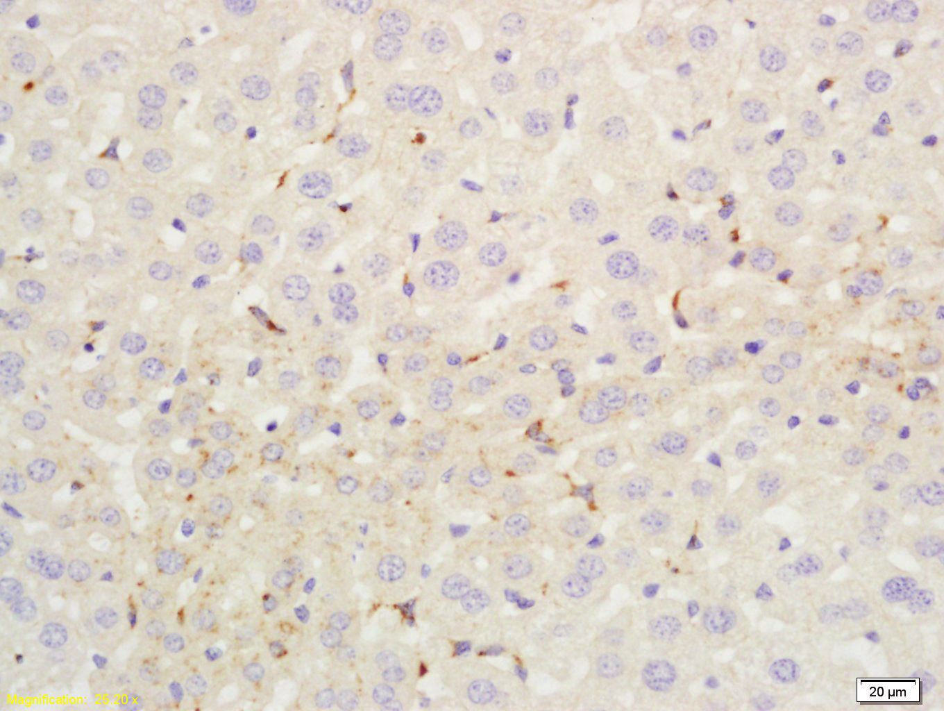
C4d-A Rabbit pAb
C4d-A Rabbit pAb
- 产品详情
- 实验流程
- 背景知识
Application
| IHC-P, IHC-F, IF |
|---|---|
| Primary Accession | P0C0L4 |
| Reactivity | Mouse |
| Predicted | Human |
| Host | Rabbit |
| Clonality | Polyclonal |
| Calculated MW | 192785 Da |
| Physical State | Liquid |
| Immunogen | KLH conjugated synthetic peptide derived from human C4d-A |
| Epitope Specificity | 1001-1100/1744 |
| Isotype | IgG |
| Purity | affinity purified by Protein A |
| Buffer | 0.01M TBS (pH7.4) with 1% BSA, 0.02% Proclin300 and 50% Glycerol. |
| SUBCELLULAR LOCATION | Secreted. |
| SIMILARITY | Contains 1 anaphylatoxin-like domain.Contains 1 NTR domain. |
| SUBUNIT | This protein is synthesized as a single-chain precursor and, prior to secretion, is enzymatically cleaved to form a trimer of non-identical chains (alpha, beta and gamma). |
| Post-translational modifications | Prior to secretion, the single-chain precursor is enzymatically cleaved to yield non-identical chains alpha, beta and gamma. During activation, the alpha chain is cleaved by C1 into C4a and C4b, and C4b stays linked to the beta and gamma chains. Further degradation of C4b by C1 into the inactive fragments C4c and C4d blocks the generation of C3 convertase. The proteolytic cleavages often are incomplete so that many structural forms can be found in plasma. N- and O-glycosylated. O-glycosylated with a core 1 or possibly core 8 glycan. |
| DISEASE | Defects in C4A are the cause of complement component 4A deficiency (C4AD). A rare defect of the complement classical pathway associated with the development of autoimmune disorders, mainly systemic lupus with or without associated glomerulonephritis. |
| Important Note | This product as supplied is intended for research use only, not for use in human, therapeutic or diagnostic applications. |
| Background Descriptions | This gene encodes the acidic form of complement factor 4, part of the classical activation pathway. The protein is expressed as a single chain precursor which is proteolytically cleaved into a trimer of alpha, beta, and gamma chains prior to secretion. The trimer provides a surface for interaction between the antigen-antibody complex and other complement components. The alpha chain may be cleaved to release C4 anaphylatoxin, a mediator of local inflammation. Deficiency of this protein is associated with systemic lupus erythematosus and type I diabetes mellitus. This gene localizes to the major histocompatibility complex (MHC) class III region on chromosome 6. Varying haplotypes of this gene cluster exist, such that individuals may have 1, 2, or 3 copies of this gene. Two transcript variants encoding different isoforms have been found for this gene. [provided by RefSeq, Nov 2011]. |
| Gene ID | 720;721 |
|---|---|
| Other Names | Complement C4-A, Acidic complement C4, C3 and PZP-like alpha-2-macroglobulin domain-containing protein 2, Complement C4 beta chain, Complement C4-A alpha chain, C4a anaphylatoxin, Complement C4b-A, Complement C4b-alpha' chain, Complement C4d-A, Complement C4 gamma chain, C4A {ECO:0000303|PubMed:6546707, ECO:0000312|HGNC:HGNC:1323} |
| Target/Specificity | Complement component C4 is expressed at highest levels in the liver, at moderate levels in the adrenal cortex, adrenal medulla, thyroid gland,and the kidney, and at lowest levels in the heart, ovary, small intestine, thymus, pancreas and spleen. The extra-hepatic sites of expression may be important for the local protection and inflammatory response. |
| Dilution | IHC-P=1:100-500,IHC-F=1:100-500,IF=1:100-500 |
| Storage | Store at -20 °C for one year. Avoid repeated freeze/thaw cycles. When reconstituted in sterile pH 7.4 0.01M PBS or diluent of antibody the antibody is stable for at least two weeks at 2-4 °C. |
| Name | C4A {ECO:0000303|PubMed:6546707, ECO:0000312|HGNC:HGNC:1323} |
|---|---|
| Function | Precursor of non-enzymatic components of the classical, lectin and GZMK complement pathways, which consist in a cascade of proteins that leads to phagocytosis and breakdown of pathogens and signaling that strengthens the adaptive immune system. |
| Cellular Location | Secreted. Synapse Cell projection, axon. Cell projection, dendrite [Complement C4b-A]: Secreted. Cell surface. Note=Covalently associated with the surface of pathogens: the internal thioester bond reacts with carbohydrate antigens on the target surface to form amide or ester bonds. |
| Tissue Location | Complement component C4 is expressed at highest levels in the liver, at moderate levels in the adrenal cortex, adrenal medulla, thyroid gland, and the kidney, and at lowest levels in the heart, ovary, small intestine, thymus, pancreas and spleen (PubMed:11367523). The extra-hepatic sites of expression may be important for the local protection and inflammatory response (PubMed:11367523). |
For Research Use Only. Not For Use In Diagnostic Procedures.
Provided below are standard protocols that you may find useful for product applications.
BACKGROUND
This gene encodes the acidic form of complement factor 4, part of the classical activation pathway. The protein is expressed as a single chain precursor which is proteolytically cleaved into a trimer of alpha, beta, and gamma chains prior to secretion. The trimer provides a surface for interaction between the antigen-antibody complex and other complement components. The alpha chain may be cleaved to release C4 anaphylatoxin, a mediator of local inflammation. Deficiency of this protein is associated with systemic lupus erythematosus and type I diabetes mellitus. This gene localizes to the major histocompatibility complex (MHC) class III region on chromosome 6. Varying haplotypes of this gene cluster exist, such that individuals may have 1, 2, or 3 copies of this gene. Two transcript variants encoding different isoforms have been found for this gene. [provided by RefSeq, Nov 2011].
终于等到您。ABCEPTA(百远生物)抗体产品。
点击下方“我要评价 ”按钮提交您的反馈信息,您的反馈和评价是我们最宝贵的财富之一,
我们将在1-3个工作日内处理您的反馈信息。
如有疑问,联系:0512-88856768 tech-china@abcepta.com.


