


 癌症的基本特征包括细胞增殖、血管生成、迁移、凋亡逃避机制和细胞永生等。找到癌症发生过程中这些通路的关键标记物和对应的抗体用于检测至关重要。
 为您推荐一个泛素化位点预测神器——泛素化分析工具,可以为您的蛋白的泛素化位点作出预测和评分。
 细胞自噬受体图形绘图工具为你的蛋白的细胞受体结合位点作出预测和评分,识别结合到自噬通路中的蛋白是非常重要的,便于让我们理解自噬在正常生理、病理过程中的作用,如发育、细胞分化、神经退化性疾病、压力条件下、感染和癌症。
Anti-PALLD Antibody
- 产品详情
- 实验流程
- 背景知识
Application
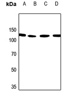
| WB |
|---|---|
| Primary Accession | Q8WX93 |
| Reactivity | Human, Mouse, Rat |
| Host | Rabbit |
| Clonality | Polyclonal |
| Calculated MW | 150564 Da |
| Gene ID | 23022 |
|---|---|
| Other Names | KIAA0992; Palladin; SIH002; Sarcoma antigen NY-SAR-77 |
| Target/Specificity | Recognizes endogenous levels of PALLD protein. |
| Dilution | WB~~1/500 - 1/1000 |
| Format | Liquid in 0.42% Potassium phosphate, 0.87% Sodium chloride, pH 7.3, 30% glycerol, and 0.09% (W/V) sodium azide. |
| Storage | Store at -20 °C.Stable for 12 months from date of receipt |
| Name | PALLD |
|---|---|
| Synonyms | KIAA0992 |
| Function | Cytoskeletal protein required for organization of normal actin cytoskeleton. Roles in establishing cell morphology, motility, cell adhesion and cell-extracellular matrix interactions in a variety of cell types. May function as a scaffolding molecule with the potential to influence both actin polymerization and the assembly of existing actin filaments into higher-order arrays. Binds to proteins that bind to either monomeric or filamentous actin. Localizes at sites where active actin remodeling takes place, such as lamellipodia and membrane ruffles. Different isoforms may have functional differences. Involved in the control of morphological and cytoskeletal changes associated with dendritic cell maturation. Involved in targeting ACTN to specific subcellular foci. |
| Cellular Location | Cytoplasm, cytoskeleton. Cell junction, focal adhesion. Cytoplasm, myofibril, sarcomere, Z line. Cell projection, ruffle. Cell projection, podosome {ECO:0000250|UniProtKB:P0C5E3}. Cell projection, lamellipodium. Cell projection, axon {ECO:0000250|UniProtKB:P0C5E3}. Cell projection, growth cone {ECO:0000250|UniProtKB:P0C5E3}. Note=Localizes to stress fibers and Z lines (PubMed:11598191, PubMed:16125169, PubMed:17322171, PubMed:17537434). Preferentially expressed in the excitatory presynaptic terminals (By similarity). {ECO:0000250|UniProtKB:P0C5E3, ECO:0000269|PubMed:11598191, ECO:0000269|PubMed:16125169, ECO:0000269|PubMed:17322171, ECO:0000269|PubMed:17537434} |
| Tissue Location | Detected in both muscle and non-muscle tissues. High expression in prostate, ovary, colon, and kidney. Not detected in spleen, skeletal muscle, lung and peripheral blood lymphocytes (at protein level). Protein is overexpressed in FA6, HPAF, IMIM-PC2, SUIT-2 and PancTu-II sporadic pancreatic cancer cell lines |
Research Areas
For Research Use Only. Not For Use In Diagnostic Procedures.
Application Protocols
Provided below are standard protocols that you may find useful for product applications.
BACKGROUND
Rabbit polyclonal antibody to PALLD
终于等到您。ABCEPTA(百远生物)抗体产品。
点击下方“我要评价 ”按钮提交您的反馈信息,您的反馈和评价是我们最宝贵的财富之一,
我们将在1-3个工作日内处理您的反馈信息。
如有疑问,联系:0512-88856768 tech-china@abcepta.com.
¥ 1,500.00
Cat# AP53784


