


 癌症的基本特征包括细胞增殖、血管生成、迁移、凋亡逃避机制和细胞永生等。找到癌症发生过程中这些通路的关键标记物和对应的抗体用于检测至关重要。
 为您推荐一个泛素化位点预测神器——泛素化分析工具,可以为您的蛋白的泛素化位点作出预测和评分。
 细胞自噬受体图形绘图工具为你的蛋白的细胞受体结合位点作出预测和评分,识别结合到自噬通路中的蛋白是非常重要的,便于让我们理解自噬在正常生理、病理过程中的作用,如发育、细胞分化、神经退化性疾病、压力条件下、感染和癌症。
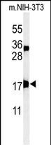
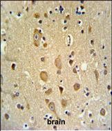
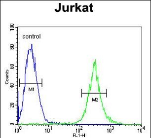
APITD1 Antibody (Center)
Affinity Purified Rabbit Polyclonal Antibody (Pab)
- 产品详情
- 实验流程
- 背景知识
Application
| WB, IHC-P, FC, E |
|---|---|
| Primary Accession | Q8N2Z9 |
| Other Accession | Q9D084, Q2TBR7 |
| Reactivity | Human, Mouse |
| Predicted | Bovine |
| Host | Rabbit |
| Clonality | Polyclonal |
| Isotype | Rabbit IgG |
| Calculated MW | 15893 Da |
| Antigen Region | 48-74 aa |
| Gene ID | 100526739;378708 |
|---|---|
| Other Names | Centromere protein S, CENP-S, Apoptosis-inducing TAF9-like domain-containing protein 1, FANCM-interacting histone fold protein 1, Fanconi anemia-associated polypeptide of 16 kDa, APITD1, CENPS, FAAP16, MHF1 |
| Target/Specificity | This APITD1 antibody is generated from rabbits immunized with a KLH conjugated synthetic peptide between 48-74 amino acids from the Central region of human APITD1. |
| Dilution | WB~~1:1000 IHC-P~~1:100~500 FC~~1:10~50 E~~Use at an assay dependent concentration. |
| Format | Purified polyclonal antibody supplied in PBS with 0.09% (W/V) sodium azide. This antibody is purified through a protein A column, followed by peptide affinity purification. |
| Storage | Maintain refrigerated at 2-8°C for up to 2 weeks. For long term storage store at -20°C in small aliquots to prevent freeze-thaw cycles. |
| Precautions | APITD1 Antibody (Center) is for research use only and not for use in diagnostic or therapeutic procedures. |
| Name | CENPS |
|---|---|
| Function | DNA-binding component of the Fanconi anemia (FA) core complex. Required for the normal activation of the FA pathway, leading to monoubiquitination of the FANCI-FANCD2 complex in response to DNA damage, cellular resistance to DNA cross-linking drugs, and prevention of chromosomal breakage (PubMed:20347428, PubMed:20347429). In complex with CENPX (MHF heterodimer), crucial cofactor for FANCM in both binding and ATP-dependent remodeling of DNA. Stabilizes FANCM (PubMed:20347428, PubMed:20347429). In complex with CENPX and FANCM (but not other FANC proteins), rapidly recruited to blocked forks and promotes gene conversion at blocked replication forks (PubMed:20347428). In complex with CENPT, CENPW and CENPX (CENP-T-W-S-X heterotetramer), involved in the formation of a functional kinetochore outer plate, which is essential for kinetochore-microtubule attachment and faithful mitotic progression (PubMed:19620631). As a component of MHF and CENP-T-W-S-X complexes, binds DNA and bends it to form a nucleosome-like structure (PubMed:20347428, PubMed:22304917). DNA- binding function is fulfilled in the presence of CENPX, with the following preference for DNA substates: Holliday junction > double- stranded > splay arm > single-stranded. Does not bind DNA on its own (PubMed:20347428, PubMed:20347429). |
| Cellular Location | Nucleus. Chromosome, centromere Chromosome, centromere, kinetochore Note=Assembly of CENPS and CENPX and its partner subunits CENPT and CENPW at centromeres occurs through a dynamic exchange mechanism Although exchange is continuous in the cell cycle, de novo assembly starts principally during mid-late S phase and is complete by G2. CENPS is more stably bound at the kinetochore than CENPX (PubMed:19620631, PubMed:24522885). During S phase, rapidly recruited to DNA interstrand cross-links that block replication (PubMed:20347428). Recruited to DNA damage sites about 20 minutes following UV irradiation, reaching a plateau after approximately 40 minutes (PubMed:24522885) |
| Tissue Location | Ubiquitously expressed. |
For Research Use Only. Not For Use In Diagnostic Procedures.
Provided below are standard protocols that you may find useful for product applications.
BACKGROUND
APITD1 was identified in the neuroblastoma tumour suppressor candidate region on chromosome 1p36. It contains a TFIID-31 domain, similar to that found in TATA box-binding protein-associated factor, TAF(II)31, which is required for p53-mediated transcription activation. This gene was expressed at very low levels in neuroblastoma tumours, and was shown to reduce cell growth in neuroblastoma cells, suggesting that it may have a role in a cell death pathway.
REFERENCES
Amano, M., et al. J. Cell Biol. 186(2):173-182(2009)
van Gils, W., et al. Invest. Ophthalmol. Vis. Sci. 48(11):4919-4923(2007)
Okada, M., et al. Nat. Cell Biol. 8(5):446-457(2006)
终于等到您。ABCEPTA(百远生物)抗体产品。
点击下方“我要评价 ”按钮提交您的反馈信息,您的反馈和评价是我们最宝贵的财富之一,
我们将在1-3个工作日内处理您的反馈信息。
如有疑问,联系:0512-88856768 tech-china@abcepta.com.


