


 癌症的基本特征包括细胞增殖、血管生成、迁移、凋亡逃避机制和细胞永生等。找到癌症发生过程中这些通路的关键标记物和对应的抗体用于检测至关重要。
 为您推荐一个泛素化位点预测神器——泛素化分析工具,可以为您的蛋白的泛素化位点作出预测和评分。
 细胞自噬受体图形绘图工具为你的蛋白的细胞受体结合位点作出预测和评分,识别结合到自噬通路中的蛋白是非常重要的,便于让我们理解自噬在正常生理、病理过程中的作用,如发育、细胞分化、神经退化性疾病、压力条件下、感染和癌症。
DBN1 Antibody (Center)
Purified Rabbit Polyclonal Antibody (Pab)
- 产品详情
- 实验流程
- 背景知识
Application
| WB, E |
|---|---|
| Primary Accession | Q16643 |
| Other Accession | Q9QXS6, Q07266 |
| Reactivity | Human, Mouse, Rat |
| Host | Rabbit |
| Clonality | polyclonal |
| Isotype | Rabbit IgG |
| Calculated MW | 71429 Da |
| Gene ID | 1627 |
|---|---|
| Other Names | Drebrin, Developmentally-regulated brain protein, DBN1, D0S117E |
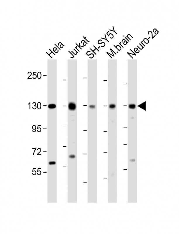
| Target/Specificity | This DBN1 antibody is generated from a rabbit immunized with a KLH conjugated synthetic peptide between 194-228 amino acids from the Central region of human DBN1. |
| Dilution | WB~~1:2000 E~~Use at an assay dependent concentration. |
| Format | Purified polyclonal antibody supplied in PBS with 0.09% (W/V) sodium azide. This antibody is purified through a protein A column, followed by peptide affinity purification. |
| Storage | Maintain refrigerated at 2-8°C for up to 2 weeks. For long term storage store at -20°C in small aliquots to prevent freeze-thaw cycles. |
| Precautions | DBN1 Antibody (Center) is for research use only and not for use in diagnostic or therapeutic procedures. |
| Name | DBN1 |
|---|---|
| Synonyms | D0S117E |
| Function | Actin cytoskeleton-organizing protein that plays a role in the formation of cell projections (PubMed:20215400). Required for actin polymerization at immunological synapses (IS) and for the recruitment of the chemokine receptor CXCR4 to IS (PubMed:20215400). Plays a role in dendritic spine morphogenesis and organization, including the localization of the dopamine receptor DRD1 to the dendritic spines (By similarity). Involved in memory-related synaptic plasticity in the hippocampus (By similarity). |
| Cellular Location | Cytoplasm. Cell projection, dendrite. Cytoplasm, cell cortex. Cell junction. Cell projection, growth cone {ECO:0000250|UniProtKB:Q9QXS6}. Note=In the absence of antigen, evenly distributed throughout subcortical regions of the T-cell membrane and cytoplasm (PubMed:20215400). In the presence of antigen, distributes to the immunological synapse forming at the T-cell-APC contact area, where it localizes at the peripheral and distal supramolecular activation clusters (SMAC) (PubMed:20215400). Colocalized with RUFY3 and F-actin at the transitional domain of the axonal growth cone (By similarity) {ECO:0000250|UniProtKB:Q9QXS6, ECO:0000269|PubMed:20215400} |
| Tissue Location | Expressed in the brain, with expression in the molecular layer of the dentate gyrus, stratum pyramidale, and stratum radiatum of the hippocampus (at protein level) (PubMed:8838578). Also expressed in the terminal varicosities distributed along dendritic trees of pyramidal cells in CA4 and CA3 of the hippocampus (at protein level) (PubMed:8838578). Expressed in pyramidal cells in CA2, CA1 and the subiculum of the hippocampus (at protein level) (PubMed:8838578) Expressed in peripheral blood lymphocytes, including T-cells (at protein level) (PubMed:20215400). Expressed in the brain (PubMed:8216329, Ref.2). Expressed in the heart, placenta, lung, skeletal muscle, kidney, pancreas, skin fibroblasts, gingival fibroblasts and bone-derived cells (Ref.2) {ECO:0000269|PubMed:20215400, ECO:0000269|PubMed:8216329, ECO:0000269|PubMed:8838578, ECO:0000269|Ref.2} |
For Research Use Only. Not For Use In Diagnostic Procedures.
Provided below are standard protocols that you may find useful for product applications.
BACKGROUND
Drebrins might play some role in cell migration, extension of neuronal processes and plasticity of dendrites. Required for actin polymerization at immunological synapses (IS) and for CXCR4 recruitment to IS.
REFERENCES
Toda M.,et al.Biochem. Biophys. Res. Commun. 196:468-472(1993).
Fisher L.W.,et al.Neurosci. Res. Commun. 14:35-42(1994).
Ota T.,et al.Nat. Genet. 36:40-45(2004).
Bechtel S.,et al.BMC Genomics 8:399-399(2007).
Schmutz J.,et al.Nature 431:268-274(2004).
终于等到您。ABCEPTA(百远生物)抗体产品。
点击下方“我要评价 ”按钮提交您的反馈信息,您的反馈和评价是我们最宝贵的财富之一,
我们将在1-3个工作日内处理您的反馈信息。
如有疑问,联系:0512-88856768 tech-china@abcepta.com.


