


 癌症的基本特征包括细胞增殖、血管生成、迁移、凋亡逃避机制和细胞永生等。找到癌症发生过程中这些通路的关键标记物和对应的抗体用于检测至关重要。
 为您推荐一个泛素化位点预测神器——泛素化分析工具,可以为您的蛋白的泛素化位点作出预测和评分。
 细胞自噬受体图形绘图工具为你的蛋白的细胞受体结合位点作出预测和评分,识别结合到自噬通路中的蛋白是非常重要的,便于让我们理解自噬在正常生理、病理过程中的作用,如发育、细胞分化、神经退化性疾病、压力条件下、感染和癌症。
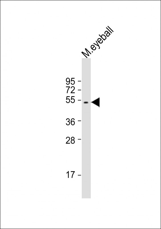
SAG Antibody (C-Term)
Purified Rabbit Polyclonal Antibody (Pab)
- 产品详情
- 实验流程
- 背景知识
Application
| WB, E |
|---|---|
| Primary Accession | P10523 |
| Reactivity | Human, Rat, Mouse |
| Host | Rabbit |
| Clonality | polyclonal |
| Isotype | Rabbit IgG |
| Calculated MW | 45120 Da |
| Gene ID | 6295 |
|---|---|
| Other Names | S-arrestin, 48 kDa protein, Retinal S-antigen, S-AG, Rod photoreceptor arrestin, SAG |
| Target/Specificity | This SAG antibody is generated from a rabbit immunized with a KLH conjugated synthetic peptide between 273-307 amino acids from the human SAG. |
| Dilution | WB~~1:2000 E~~Use at an assay dependent concentration. |
| Format | Purified polyclonal antibody supplied in PBS with 0.09% (W/V) sodium azide. This antibody is purified through a protein A column, followed by peptide affinity purification. |
| Storage | Maintain refrigerated at 2-8°C for up to 2 weeks. For long term storage store at -20°C in small aliquots to prevent freeze-thaw cycles. |
| Precautions | SAG Antibody (C-Term) is for research use only and not for use in diagnostic or therapeutic procedures. |
| Name | SAG |
|---|---|
| Function | Binds to photoactivated, phosphorylated RHO and terminates RHO signaling via G-proteins by competing with G-proteins for the same binding site on RHO (By similarity). May play a role in preventing light-dependent degeneration of retinal photoreceptor cells (PubMed:9565049). |
| Cellular Location | Cell projection, cilium, photoreceptor outer segment. Membrane {ECO:0000250|UniProtKB:P20443}; Peripheral membrane protein {ECO:0000250|UniProtKB:P20443}. Note=Highly expressed in photoreceptor outer segments in light-exposed retina. Evenly distributed throughout rod photoreceptor cells in dark-adapted retina (By similarity) Predominantly dectected at the proximal region of photoreceptor outer segments, near disk membranes (PubMed:3720866) {ECO:0000250|UniProtKB:P08168, ECO:0000269|PubMed:3720866} |
| Tissue Location | Detected in retina, in the proximal portion of the outer segment of rod photoreceptor cells (at protein level) |
For Research Use Only. Not For Use In Diagnostic Procedures.
Provided below are standard protocols that you may find useful for product applications.
BACKGROUND
Arrestin is one of the major proteins of the ros (retinal rod outer segments); it binds to photoactivated- phosphorylated rhodopsin, thereby apparently preventing the transducin-mediated activation of phosphodiesterase.
REFERENCES
Yamaki K.,et al.FEBS Lett. 234:39-43(1988).
Yamaki K.,et al.FEBS Lett. 236:507-507(1988).
Yamamoto S.,et al.Nat. Genet. 15:175-178(1997).
Hillier L.W.,et al.Nature 434:724-731(2005).
Roni V.,et al.BMC Genomics 8:42-42(2007).
终于等到您。ABCEPTA(百远生物)抗体产品。
点击下方“我要评价 ”按钮提交您的反馈信息,您的反馈和评价是我们最宝贵的财富之一,
我们将在1-3个工作日内处理您的反馈信息。
如有疑问,联系:0512-88856768 tech-china@abcepta.com.
|
刘飞
2015-12-07 16:03:52
1
2
3
4
5
|
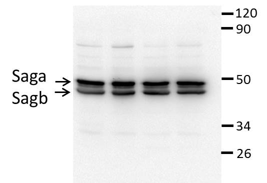
Species tested
zebrafish
Application tested
western blot
Organization tested
eyes
Barcode encoding
Brief protocol
|
|


