


 癌症的基本特征包括细胞增殖、血管生成、迁移、凋亡逃避机制和细胞永生等。找到癌症发生过程中这些通路的关键标记物和对应的抗体用于检测至关重要。
 为您推荐一个泛素化位点预测神器——泛素化分析工具,可以为您的蛋白的泛素化位点作出预测和评分。
 细胞自噬受体图形绘图工具为你的蛋白的细胞受体结合位点作出预测和评分,识别结合到自噬通路中的蛋白是非常重要的,便于让我们理解自噬在正常生理、病理过程中的作用,如发育、细胞分化、神经退化性疾病、压力条件下、感染和癌症。
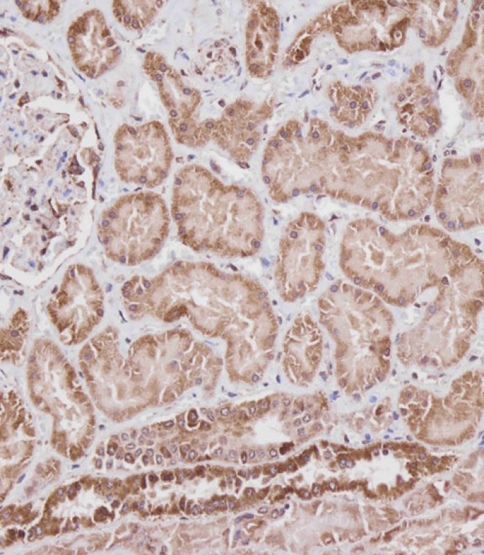
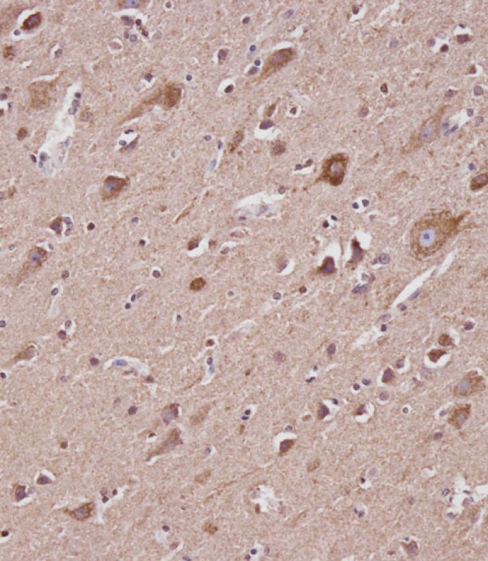
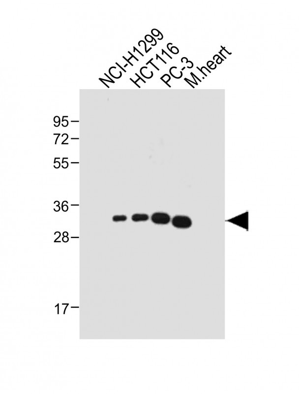
VDAC2 Antibody (N-term)
Purified Rabbit Polyclonal Antibody (Pab)
- 产品详情
- 实验流程
- 背景知识
Application
| IHC-P, WB, E |
|---|---|
| Primary Accession | P45880 |
| Reactivity | Human, Rat, Mouse |
| Host | Rabbit |
| Clonality | polyclonal |
| Isotype | Rabbit IgG |
| Calculated MW | 31567 Da |
| Gene ID | 7417 |
|---|---|
| Other Names | Voltage-dependent anion-selective channel protein 2, VDAC-2, hVDAC2, Outer mitochondrial membrane protein porin 2, VDAC2 |
| Target/Specificity | This VDAC2 antibody is generated from a rabbit immunized with a KLH conjugated synthetic peptide between 51-85 amino acids from the N-terminal region of human VDAC2. |
| Dilution | IHC-P~~1:100 WB~~1:4000 E~~Use at an assay dependent concentration. |
| Format | Purified polyclonal antibody supplied in PBS with 0.09% (W/V) sodium azide. This antibody is purified through a protein A column, followed by peptide affinity purification. |
| Storage | Maintain refrigerated at 2-8°C for up to 2 weeks. For long term storage store at -20°C in small aliquots to prevent freeze-thaw cycles. |
| Precautions | VDAC2 Antibody (N-term) is for research use only and not for use in diagnostic or therapeutic procedures. |
| Name | VDAC2 (HGNC:12672) |
|---|---|
| Function | Non-selective voltage-gated ion channel that mediates the transport of anions and cations through the mitochondrion outer membrane and plasma membrane (PubMed:8420959). The channel adopts an open conformation at zero mV and a closed conformation at both positive and negative potentials (PubMed:8420959). There are two populations of channels; the main that functions in a lower open-state conductance with lower ion selectivity, that switch, in a voltage-dependent manner, from the open to a low-conducting 'closed' state and the other that has a normal ion selectivity in the typical high conductance, 'open' state (PubMed:8420959). Binds various lipids, including the sphingolipid ceramide, the phospholipid phosphatidylcholine, and the sterols cholesterol and oxysterol (PubMed:31015432). Binding of ceramide promotes the mitochondrial outer membrane permeabilization (MOMP) apoptotic pathway (PubMed:31015432). Associates with the translocase of the outer mitochondrial membrane (TOM) complex and PINK1 kinase at depolarized mitochondria, this interaction stabilizes PINK1 at the outer mitochondrial membrane and triggers downstream mitophagy by the recruitment of the E3 ubiquitin ligase PRKN (PubMed:40080546). |
| Cellular Location | Mitochondrion outer membrane; Multi-pass membrane protein. Membrane. Note=May localize to non-mitochondrial membranes. |
| Tissue Location | Expressed in erythrocytes (at protein level) (PubMed:27641616). Expressed in all tissues examined (PubMed:8420959) |
For Research Use Only. Not For Use In Diagnostic Procedures.
Provided below are standard protocols that you may find useful for product applications.
BACKGROUND
Forms a channel through the mitochondrial outer membrane that allows diffusion of small hydrophilic molecules. The channel adopts an open conformation at low or zero membrane potential and a closed conformation at potentials above 30-40 mV. The open state has a weak anion selectivity whereas the closed state is cation- selective.
REFERENCES
Ha H.,et al.J. Biol. Chem. 268:12143-12149(1993).
Blachly-Dyson E.,et al.J. Biol. Chem. 268:1835-1841(1993).
Decker W.K.,et al.Mamm. Genome 10:1041-1042(1999).
Ebert L.,et al.Submitted (JUN-2004) to the EMBL/GenBank/DDBJ databases.
Deloukas P.,et al.Nature 429:375-381(2004).
终于等到您。ABCEPTA(百远生物)抗体产品。
点击下方“我要评价 ”按钮提交您的反馈信息,您的反馈和评价是我们最宝贵的财富之一,
我们将在1-3个工作日内处理您的反馈信息。
如有疑问,联系:0512-88856768 tech-china@abcepta.com.


