


 癌症的基本特征包括细胞增殖、血管生成、迁移、凋亡逃避机制和细胞永生等。找到癌症发生过程中这些通路的关键标记物和对应的抗体用于检测至关重要。
 为您推荐一个泛素化位点预测神器——泛素化分析工具,可以为您的蛋白的泛素化位点作出预测和评分。
 细胞自噬受体图形绘图工具为你的蛋白的细胞受体结合位点作出预测和评分,识别结合到自噬通路中的蛋白是非常重要的,便于让我们理解自噬在正常生理、病理过程中的作用,如发育、细胞分化、神经退化性疾病、压力条件下、感染和癌症。
ROCK2 Antibody (C-term)
Purified Rabbit Polyclonal Antibody (Pab)
- 产品详情
- 实验流程
- 背景知识
Application
| WB, E |
|---|---|
| Primary Accession | O75116 |
| Reactivity | Human, Rat, Mouse |
| Host | Rabbit |
| Clonality | polyclonal |
| Isotype | Rabbit IgG |
| Calculated MW | 160900 Da |
| Gene ID | 9475 |
|---|---|
| Other Names | Rho-associated protein kinase 2, Rho kinase 2, Rho-associated, coiled-coil-containing protein kinase 2, Rho-associated, coiled-coil-containing protein kinase II, ROCK-II, p164 ROCK-2, ROCK2, KIAA0619 |
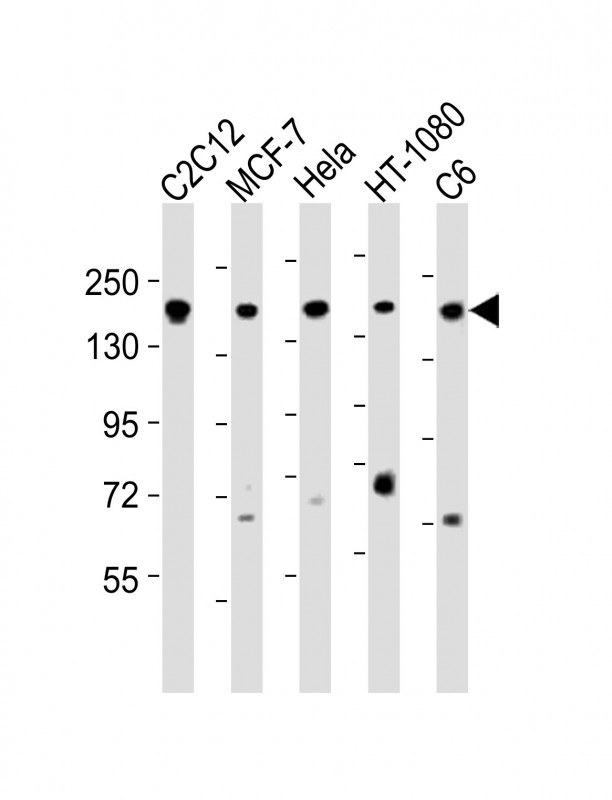
| Target/Specificity | This ROCK2 antibody is generated from a rabbit immunized with a KLH conjugated synthetic peptide between 1040-1073 amino acids from the C-terminal region of human ROCK2. |
| Dilution | WB~~1:2000 E~~Use at an assay dependent concentration. |
| Format | Purified polyclonal antibody supplied in PBS with 0.09% (W/V) sodium azide. This antibody is purified through a protein A column, followed by peptide affinity purification. |
| Storage | Maintain refrigerated at 2-8°C for up to 2 weeks. For long term storage store at -20°C in small aliquots to prevent freeze-thaw cycles. |
| Precautions | ROCK2 Antibody (C-term) is for research use only and not for use in diagnostic or therapeutic procedures. |
| Name | ROCK2 |
|---|---|
| Synonyms | KIAA0619 |
| Function | Protein kinase which is a key regulator of actin cytoskeleton and cell polarity. Involved in regulation of smooth muscle contraction, actin cytoskeleton organization, stress fiber and focal adhesion formation, neurite retraction, cell adhesion and motility via phosphorylation of ADD1, BRCA2, CNN1, EZR, DPYSL2, EP300, MSN, MYL9/MLC2, NPM1, RDX, PPP1R12A and VIM. Phosphorylates SORL1 and IRF4. Acts as a negative regulator of VEGF-induced angiogenic endothelial cell activation. Positively regulates the activation of p42/MAPK1- p44/MAPK3 and of p90RSK/RPS6KA1 during myogenic differentiation. Plays an important role in the timely initiation of centrosome duplication. Inhibits keratinocyte terminal differentiation. May regulate closure of the eyelids and ventral body wall through organization of actomyosin bundles. Plays a critical role in the regulation of spine and synaptic properties in the hippocampus. Plays an important role in generating the circadian rhythm of the aortic myofilament Ca(2+) sensitivity and vascular contractility by modulating the myosin light chain phosphorylation. |
| Cellular Location | Cytoplasm. Cell membrane; Peripheral membrane protein. Nucleus. Cytoplasm, cytoskeleton, microtubule organizing center, centrosome Note=Cytoplasmic, and associated with actin microfilaments and the plasma membrane. |
| Tissue Location | Expressed in the brain (at protein level). |
For Research Use Only. Not For Use In Diagnostic Procedures.
Provided below are standard protocols that you may find useful for product applications.
BACKGROUND
Protein kinase which is a key regulator of actin cytoskeleton and cell polarity. Involved in regulation of smooth muscle contraction, actin cytoskeleton organization, stress fiber and focal adhesion formation, neurite retraction, cell adhesion and motility via phosphorylation of ADD1, BRCA2, CNN1, EZR, DPYSL2, EP300, MSN, MYL9/MLC2, NPM1, RDX, PPP1R12A and VIM. Phosphorylates SORL1 and IRF4. Acts as a negative regulator of VEGF-induced angiogenic endothelial cell activation. Positively regulates the activation of p42/MAPK1-p44/MAPK3 and of p90RSK/RPS6KA1 during myogenic differentiation. Plays an important role in the timely initiation of centrosome duplication. Inhibits keratinocyte terminal differentiation. May regulate closure of the eyelids and ventral body wall through organization of actomyosin bundles. Plays a critical role in the regulation of spine and synaptic properties in the hippocampus. Plays an important role in generating the circadian rhythm of the aortic myofilament Ca(2+) sensitivity and vascular contractility by modulating the myosin light chain phosphorylation.
REFERENCES
Takahashi N.,et al.Genomics 55:235-237(1999).
Ishikawa K.,et al.DNA Res. 5:169-176(1998).
Hillier L.W.,et al.Nature 434:724-731(2005).
Kawano Y.,et al.J. Cell Biol. 147:1023-1038(1999).
Sebbagh M.,et al.J. Exp. Med. 201:465-471(2005).
终于等到您。ABCEPTA(百远生物)抗体产品。
点击下方“我要评价 ”按钮提交您的反馈信息,您的反馈和评价是我们最宝贵的财富之一,
我们将在1-3个工作日内处理您的反馈信息。
如有疑问,联系:0512-88856768 tech-china@abcepta.com.


