


 癌症的基本特征包括细胞增殖、血管生成、迁移、凋亡逃避机制和细胞永生等。找到癌症发生过程中这些通路的关键标记物和对应的抗体用于检测至关重要。
 为您推荐一个泛素化位点预测神器——泛素化分析工具,可以为您的蛋白的泛素化位点作出预测和评分。
 细胞自噬受体图形绘图工具为你的蛋白的细胞受体结合位点作出预测和评分,识别结合到自噬通路中的蛋白是非常重要的,便于让我们理解自噬在正常生理、病理过程中的作用,如发育、细胞分化、神经退化性疾病、压力条件下、感染和癌症。
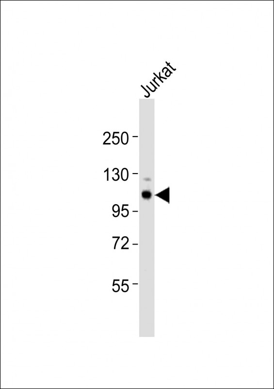
(Mouse) Uhrf2 Antibody (Center)
Purified Rabbit Polyclonal Antibody (Pab)
- 产品详情
- 实验流程
- 背景知识
Application
| WB, E |
|---|---|
| Primary Accession | Q7TMI3 |
| Reactivity | Human, Mouse |
| Host | Rabbit |
| Clonality | polyclonal |
| Isotype | Rabbit IgG |
| Calculated MW | 90106 Da |
| Gene ID | 109113 |
|---|---|
| Other Names | E3 ubiquitin-protein ligase UHRF2, 632-, NIRF, Np95-like ring finger protein, Nuclear protein 97, Nuclear zinc finger protein Np97, Ubiquitin-like PHD and RING finger domain-containing protein 2, Ubiquitin-like-containing PHD and RING finger domains protein 2, Uhrf2, Nirf |
| Target/Specificity | This Mouse Uhrf2 antibody is generated from a rabbit immunized with a KLH conjugated synthetic peptide between 472-505 amino acids from the Central region of Mouse Uhrf2. |
| Dilution | WB~~1:1000 E~~Use at an assay dependent concentration. |
| Format | Purified polyclonal antibody supplied in PBS with 0.09% (W/V) sodium azide. This antibody is purified through a protein A column, followed by peptide affinity purification. |
| Storage | Maintain refrigerated at 2-8°C for up to 2 weeks. For long term storage store at -20°C in small aliquots to prevent freeze-thaw cycles. |
| Precautions | (Mouse) Uhrf2 Antibody (Center) is for research use only and not for use in diagnostic or therapeutic procedures. |
| Name | Uhrf2 |
|---|---|
| Synonyms | Nirf |
| Function | E3 ubiquitin ligase that plays important roles in DNA methylation, histone modifications, cell cycle and DNA repair. Acts as a specific reader for 5-hydroxymethylcytosine (5hmC) and thereby recruits various substrates to these sites to ubiquitinate them (PubMed:23434322, PubMed:28402695). This activity also allows the maintenance of 5mC levels at specific genomic loci and regulates neuron-related gene expression (PubMed:28115522). Participates in cell cycle regulation by ubiquitinating cyclins CCND1 and CCNE1 and thus inducing G1 arrest. Also ubiquitinates PCNP leading to its degradation by the proteasome. Plays an active role in DNA damage repair by ubiquitinating p21/CDKN1A leading to its proteasomal degradation. Also promotes DNA repair by acting as an interstrand cross-links (ICLs) sensor. Mechanistically, cooperates with UHRF1 to ensure recruitment of FANCD2 to ICLs, leading to FANCD2 monoubiquitination and subsequent activation. Contributes to UV-induced DNA damage response by physically interacting with ATR in response to irradiation, thereby promoting ATR activation (By similarity). |
| Cellular Location | Nucleus {ECO:0000255|PROSITE-ProRule:PRU00358, ECO:0000269|PubMed:21598301}. Chromosome {ECO:0000250|UniProtKB:Q96PU4}. Note=Enriched at genomic loci that are enriched for 5-hydroxy-methylcytosine (5hmC) {ECO:0000250|UniProtKB:Q96PU4} |
| Tissue Location | Mostly detected in several tissues, including the thymus, spleen, lung, adrenal gland, and ovary. In addition, found in several tissues in the brain (cerebellum, hippocampus, and cerebral cortex). |
For Research Use Only. Not For Use In Diagnostic Procedures.
Provided below are standard protocols that you may find useful for product applications.
BACKGROUND
E3 SUMO-, but not ubiquitin-, protein ligase for ZNF131 (By similarity). E3 ubiquitin-protein ligase that is an intermolecular hub protein in the cell cycle network. Ubiquitinates cyclins, CCND1 and CCNE1, in an apparently phosphorylation-independent manner and induces G1 arrest. Also ubiquitinates PCNP leading to its degradation by the proteasome. Through cooperative DNA and histone binding, may contribute to a tighter epigenetic control of gene expression in differentiated cells.
REFERENCES
Davenport J.W.,et al.Submitted (JUN-2000) to the EMBL/GenBank/DDBJ databases.
Mori T.,et al.Submitted (AUG-2003) to the EMBL/GenBank/DDBJ databases.
Carninci P.,et al.Science 309:1559-1563(2005).
Pichler G.,et al.J. Cell. Biochem. 112:2585-2593(2011).
终于等到您。ABCEPTA(百远生物)抗体产品。
点击下方“我要评价 ”按钮提交您的反馈信息,您的反馈和评价是我们最宝贵的财富之一,
我们将在1-3个工作日内处理您的反馈信息。
如有疑问,联系:0512-88856768 tech-china@abcepta.com.


