


 癌症的基本特征包括细胞增殖、血管生成、迁移、凋亡逃避机制和细胞永生等。找到癌症发生过程中这些通路的关键标记物和对应的抗体用于检测至关重要。
 为您推荐一个泛素化位点预测神器——泛素化分析工具,可以为您的蛋白的泛素化位点作出预测和评分。
 细胞自噬受体图形绘图工具为你的蛋白的细胞受体结合位点作出预测和评分,识别结合到自噬通路中的蛋白是非常重要的,便于让我们理解自噬在正常生理、病理过程中的作用,如发育、细胞分化、神经退化性疾病、压力条件下、感染和癌症。
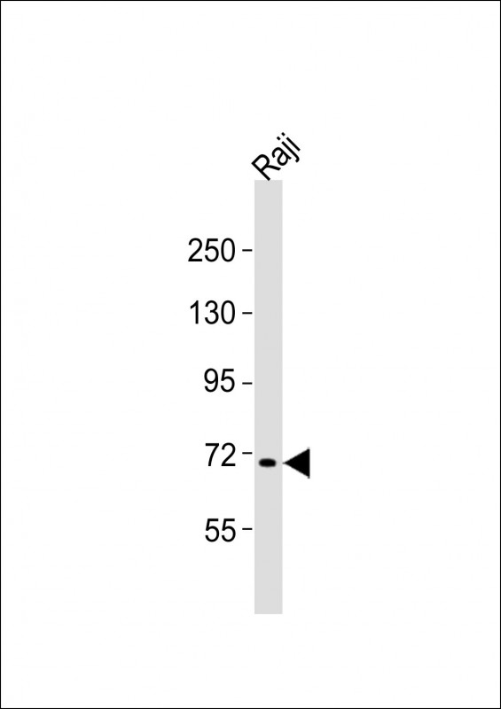
FBXW7 Antibody (N-term)
Purified Rabbit Polyclonal Antibody (Pab)
- 产品详情
- 实验流程
- 背景知识
Application
| WB, E |
|---|---|
| Primary Accession | Q969H0 |
| Reactivity | Human, Mouse |
| Host | Rabbit |
| Clonality | polyclonal |
| Isotype | Rabbit IgG |
| Calculated MW | 79663 Da |
| Gene ID | 55294 |
|---|---|
| Other Names | F-box/WD repeat-containing protein 7, Archipelago homolog, hAgo, F-box and WD-40 domain-containing protein 7, F-box protein FBX30, SEL-10, hCdc4, FBXW7 (HGNC:16712) |
| Target/Specificity | This FBXW7 antibody is generated from a rabbit immunized with a KLH conjugated synthetic peptide between 177-208 amino acids from the N-terminal region of human FBXW7. |
| Dilution | WB~~1:2000 E~~Use at an assay dependent concentration. |
| Format | Purified polyclonal antibody supplied in PBS with 0.09% (W/V) sodium azide. This antibody is purified through a protein A column, followed by peptide affinity purification. |
| Storage | Maintain refrigerated at 2-8°C for up to 2 weeks. For long term storage store at -20°C in small aliquots to prevent freeze-thaw cycles. |
| Precautions | FBXW7 Antibody (N-term) is for research use only and not for use in diagnostic or therapeutic procedures. |
| Name | FBXW7 (HGNC:16712) |
|---|---|
| Function | Substrate recognition component of a SCF (SKP1-CUL1-F-box protein) E3 ubiquitin-protein ligase complex which mediates the ubiquitination and subsequent proteasomal degradation of target proteins (PubMed:17434132, PubMed:22748924, PubMed:26976582, PubMed:28727686, PubMed:34741373, PubMed:35395208). Recognizes and binds phosphorylated sites/phosphodegrons within target proteins and thereafter brings them to the SCF complex for ubiquitination (PubMed:17434132, PubMed:22748924, PubMed:26774286, PubMed:26976582, PubMed:28727686, PubMed:34741373). Identified substrates include cyclin-E (CCNE1 or CCNE2), DISC1, JUN, MYC, NOTCH1 released notch intracellular domain (NICD), NFE2L1, NOTCH2, MCL1, MLST8, RICTOR, and probably PSEN1 (PubMed:11565034, PubMed:11585921, PubMed:12354302, PubMed:14739463, PubMed:15103331, PubMed:17558397, PubMed:17873522, PubMed:22608923, PubMed:22748924, PubMed:25775507, PubMed:25897075, PubMed:26976582, PubMed:28007894, PubMed:28727686, PubMed:29149593, PubMed:34102342). Acts as a negative regulator of JNK signaling by binding to phosphorylated JUN and promoting its ubiquitination and subsequent degradation (PubMed:14739463). Involved in bone homeostasis and negative regulation of osteoclast differentiation (PubMed:29149593). Regulates the amplitude of the cyclic expression of hepatic core clock genes and genes involved in lipid and glucose metabolism via ubiquitination and proteasomal degradation of their transcriptional repressor NR1D1; CDK1-dependent phosphorylation of NR1D1 is necessary for SCF(FBXW7)-mediated ubiquitination (PubMed:27238018). Also able to promote 'Lys-63'-linked ubiquitination in response to DNA damage (PubMed:26774286). The SCF(FBXW7) complex facilitates double-strand break repair following phosphorylation by ATM: phosphorylation promotes localization to sites of double-strand breaks and 'Lys-63'-linked ubiquitination of phosphorylated XRCC4, enhancing DNA non-homologous end joining (PubMed:26774286). |
| Cellular Location | [Isoform 1]: Nucleus, nucleoplasm. Chromosome Note=Localizes to site of double-strand breaks following phosphorylation by ATM. [Isoform 3]: Nucleus, nucleolus |
| Tissue Location | [Isoform 1]: Widely expressed. |
For Research Use Only. Not For Use In Diagnostic Procedures.
Provided below are standard protocols that you may find useful for product applications.
BACKGROUND
Substrate recognition component of an SCF (SKP1-CUL1-F- box protein) E3 ubiquitin-protein ligase complex which mediates the ubiquitination and subsequent proteasomal degradation of target proteins. Recognizes and binds phosphorylated sites/phosphodegrons within target proteins and thereafter bring them to the SCF complex for ubiquitination. Identified substrates include cyclin-E, MYC, NOTCH1 released notch intracellular domain (NICD), and probably PSEN1.
REFERENCES
Winston J.T.,et al.Curr. Biol. 9:1180-1182(1999).
Moberg K.H.,et al.Nature 413:311-316(2001).
Strohmaier H.,et al.Nature 413:316-322(2001).
Li J.,et al.J. Neurochem. 82:1540-1548(2002).
Bechtel S.,et al.BMC Genomics 8:399-399(2007).
终于等到您。ABCEPTA(百远生物)抗体产品。
点击下方“我要评价 ”按钮提交您的反馈信息,您的反馈和评价是我们最宝贵的财富之一,
我们将在1-3个工作日内处理您的反馈信息。
如有疑问,联系:0512-88856768 tech-china@abcepta.com.


