


 癌症的基本特征包括细胞增殖、血管生成、迁移、凋亡逃避机制和细胞永生等。找到癌症发生过程中这些通路的关键标记物和对应的抗体用于检测至关重要。
 为您推荐一个泛素化位点预测神器——泛素化分析工具,可以为您的蛋白的泛素化位点作出预测和评分。
 细胞自噬受体图形绘图工具为你的蛋白的细胞受体结合位点作出预测和评分,识别结合到自噬通路中的蛋白是非常重要的,便于让我们理解自噬在正常生理、病理过程中的作用,如发育、细胞分化、神经退化性疾病、压力条件下、感染和癌症。
ADRA1B Antibody (C-term)
Affinity Purified Rabbit Polyclonal Antibody (Pab)
- 产品详情
- 实验流程
- 背景知识
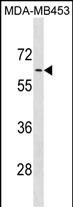
Application
| WB, E |
|---|---|
| Primary Accession | P35368 |
| Other Accession | P15823, P97717, NP_000670.1 |
| Reactivity | Human |
| Predicted | Mouse, Rat |
| Host | Rabbit |
| Clonality | Polyclonal |
| Isotype | Rabbit IgG |
| Calculated MW | 56836 Da |
| Antigen Region | 380-409 aa |
| Gene ID | 147 |
|---|---|
| Other Names | Alpha-1B adrenergic receptor, Alpha-1B adrenoreceptor, Alpha-1B adrenoceptor, ADRA1B |
| Target/Specificity | This ADRA1B antibody is generated from rabbits immunized with a KLH conjugated synthetic peptide between 380-409 amino acids from the C-terminal region of human ADRA1B. |
| Dilution | WB~~1:1000 E~~Use at an assay dependent concentration. |
| Format | Purified polyclonal antibody supplied in PBS with 0.09% (W/V) sodium azide. This antibody is purified through a protein A column, followed by peptide affinity purification. |
| Storage | Maintain refrigerated at 2-8°C for up to 2 weeks. For long term storage store at -20°C in small aliquots to prevent freeze-thaw cycles. |
| Precautions | ADRA1B Antibody (C-term) is for research use only and not for use in diagnostic or therapeutic procedures. |
| Name | ADRA1B (HGNC:278) |
|---|---|
| Function | Alpha-1 adrenergic receptors are G protein-coupled receptors for catecholamines that signal through the G(q) family of G proteins, including G(q) and G(11). Upon activation, they stimulate the phosphatidylinositol-calcium second messenger pathway, leading to calcium release from intracellular stores and activation of protein kinase C (By similarity). ADRA1B binds the catecholamine ligands norepinephrine and epinephrine (PubMed:7815325, PubMed:8183249). Can also couple to G(14) and G(16) proteins (By similarity). Nuclear ADRA1B forms heterooligomers with ADRA1A to regulate phenylephrine(PE)- stimulated ERK signaling in cardiac myocytes (PubMed:18802028, PubMed:22120526). At the plasma membrane, ADRA1B interacts with CAVIN4/MURC to regulates ERK activation in cardiomyocytes, contributing to the regulation of cardiac hypertrophy (PubMed:24567387). |
| Cellular Location | Nucleus membrane; Multi-pass membrane protein. Cell membrane; Multi-pass membrane protein. Cytoplasm. Membrane, caveola. Note=Location at the nuclear membrane facilitates heterooligomerization and regulates ERK-mediated signaling in cardiac myocytes. Colocalizes with GNAQ, PLCB1 as well as LAP2 at the nuclear membrane of cardiac myocytes (PubMed:18802028, PubMed:22120526). Colocalizes with CAVIN4 and CAV3 at the plasma membrane and partly within the cytoplasm in cardiomyocytes (PubMed:24567387). |
For Research Use Only. Not For Use In Diagnostic Procedures.
Provided below are standard protocols that you may find useful for product applications.
BACKGROUND
Alpha-1-adrenergic receptors (alpha-1-ARs) are members of the G protein-coupled receptor superfamily. They activate mitogenic responses and regulate growth and proliferation of many cells. There are 3 alpha-1-AR subtypes: alpha-1A, -1B and -1D, all of which signal through the Gq/11 family of G-proteins and different subtypes show different patterns of activation. This gene encodes alpha-1B-adrenergic receptor, which induces neoplastic transformation when transfected into NIH 3T3 fibroblasts and other cell lines. Thus, this normal cellular gene is identified as a protooncogene. This gene comprises 2 exons and a single large intron of at least 20 kb that interrupts the coding region.
REFERENCES
Bailey, S.D., et al. Diabetes Care 33(10):2250-2253(2010)
Pinheiro, A.P., et al. Am. J. Med. Genet. B Neuropsychiatr. Genet. 153B (5), 1070-1080 (2010) :
Rose, J.E., et al. Mol. Med. 16 (7-8), 247-253 (2010) :
Jensen, B.C., et al. Circ Heart Fail 2(6):654-663(2009)
Talmud, P.J., et al. Am. J. Hum. Genet. 85(5):628-642(2009)
终于等到您。ABCEPTA(百远生物)抗体产品。
点击下方“我要评价 ”按钮提交您的反馈信息,您的反馈和评价是我们最宝贵的财富之一,
我们将在1-3个工作日内处理您的反馈信息。
如有疑问,联系:0512-88856768 tech-china@abcepta.com.


