


 癌症的基本特征包括细胞增殖、血管生成、迁移、凋亡逃避机制和细胞永生等。找到癌症发生过程中这些通路的关键标记物和对应的抗体用于检测至关重要。
 为您推荐一个泛素化位点预测神器——泛素化分析工具,可以为您的蛋白的泛素化位点作出预测和评分。
 细胞自噬受体图形绘图工具为你的蛋白的细胞受体结合位点作出预测和评分,识别结合到自噬通路中的蛋白是非常重要的,便于让我们理解自噬在正常生理、病理过程中的作用,如发育、细胞分化、神经退化性疾病、压力条件下、感染和癌症。
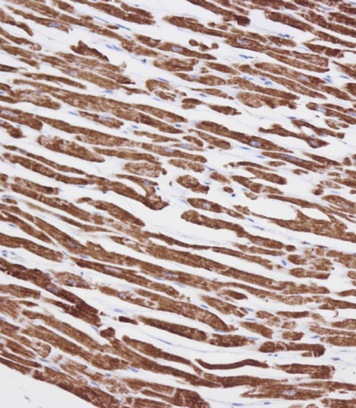
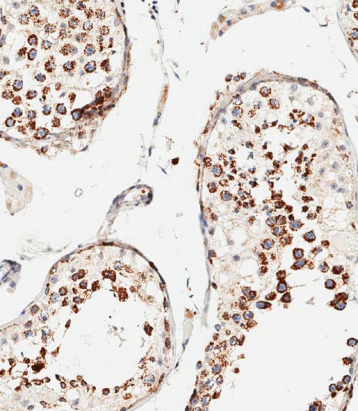
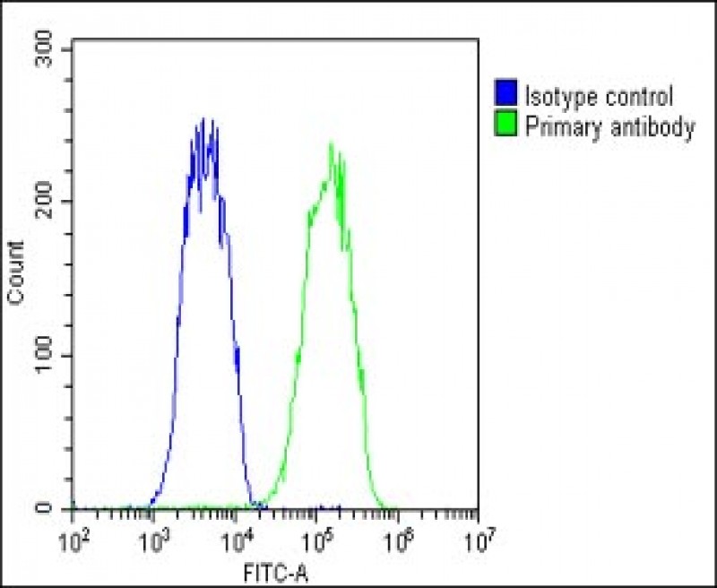
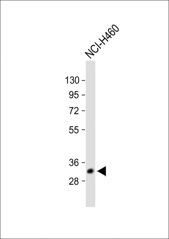
VDAC3 Antibody (Center)
Affinity Purified Rabbit Polyclonal Antibody (Pab)
- 产品详情
- 文献引用 : 1
- 实验流程
- 背景知识
Application
| IHC-P, WB, FC, IHC-P-Leica, E |
|---|---|
| Primary Accession | Q9Y277 |
| Other Accession | Q9TT13, Q29380, Q60931, Q9MZ13, NP_005653.3, NP_001129166.1 |
| Reactivity | Human, Rat, Mouse |
| Predicted | Bovine, Mouse, Pig, Rabbit |
| Host | Rabbit |
| Clonality | Polyclonal |
| Isotype | Rabbit IgG |
| Calculated MW | 30659 Da |
| Antigen Region | 156-183 aa |
| Gene ID | 7419 |
|---|---|
| Other Names | Voltage-dependent anion-selective channel protein 3, VDAC-3, hVDAC3, Outer mitochondrial membrane protein porin 3, VDAC3 |
| Target/Specificity | This VDAC3 antibody is generated from rabbits immunized with a KLH conjugated synthetic peptide between 156-183 amino acids from the Central region of human VDAC3. |
| Dilution | IHC-P~~1:100 WB~~1:1000 FC~~1:25 IHC-P-Leica~~1:500 E~~Use at an assay dependent concentration. |
| Format | Purified polyclonal antibody supplied in PBS with 0.09% (W/V) sodium azide. This antibody is purified through a protein A column, followed by peptide affinity purification. |
| Storage | Maintain refrigerated at 2-8°C for up to 2 weeks. For long term storage store at -20°C in small aliquots to prevent freeze-thaw cycles. |
| Precautions | VDAC3 Antibody (Center) is for research use only and not for use in diagnostic or therapeutic procedures. |
| Name | VDAC3 (HGNC:12674) |
|---|---|
| Function | Non-selective voltage-gated ion channel that mediates the transport of anions and cations through the mitochondrion outer membrane and plasma membrane (PubMed:31935282). Forms a high-conducting channel with a stable open state and a voltage-induced closure with a mild preference for anions over cations (PubMed:31935282). Involved in male fertility and sperm mitochondrial sheath formation (By similarity). |
| Cellular Location | Mitochondrion outer membrane {ECO:0000250|UniProtKB:P21796}. Membrane Note=May localize to non-mitochondrial membranes |
| Tissue Location | Expressed in erythrocytes (at protein level) (PubMed:27641616). Widely expressed. Highest in testis (PubMed:9781040). |
For Research Use Only. Not For Use In Diagnostic Procedures.

Provided below are standard protocols that you may find useful for product applications.
BACKGROUND
VDAC3 belongs to a group of mitochondrial membrane channels involved in translocation of adenine nucleotides through the outer membrane. These channels may also function as a mitochondrial binding site for hexokinase (see HK1; MIM 142600) and glycerol kinase (GK; MIM 300474) (Rahmani et al., 1998).[supplied by OMIM].
REFERENCES
Reina, S., et al. FEBS Lett. 584(13):2837-2844(2010)
Lefievre, L., et al. Proteomics 7(17):3066-3084(2007)
Lamesch, P., et al. Genomics 89(3):307-315(2007)
Ewing, R.M., et al. Mol. Syst. Biol. 3, 89 (2007) :
Rush, J., et al. Nat. Biotechnol. 23(1):94-101(2005)
终于等到您。ABCEPTA(百远生物)抗体产品。
点击下方“我要评价 ”按钮提交您的反馈信息,您的反馈和评价是我们最宝贵的财富之一,
我们将在1-3个工作日内处理您的反馈信息。
如有疑问,联系:0512-88856768 tech-china@abcepta.com.


