


 癌症的基本特征包括细胞增殖、血管生成、迁移、凋亡逃避机制和细胞永生等。找到癌症发生过程中这些通路的关键标记物和对应的抗体用于检测至关重要。
 为您推荐一个泛素化位点预测神器——泛素化分析工具,可以为您的蛋白的泛素化位点作出预测和评分。
 细胞自噬受体图形绘图工具为你的蛋白的细胞受体结合位点作出预测和评分,识别结合到自噬通路中的蛋白是非常重要的,便于让我们理解自噬在正常生理、病理过程中的作用,如发育、细胞分化、神经退化性疾病、压力条件下、感染和癌症。
RRH Antibody (C-term)
Affinity Purified Rabbit Polyclonal Antibody (Pab)
- 产品详情
- 实验流程
- 背景知识
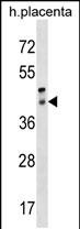
Application
| WB, E |
|---|---|
| Primary Accession | O14718 |
| Other Accession | NP_006574.1 |
| Reactivity | Human |
| Host | Rabbit |
| Clonality | Polyclonal |
| Isotype | Rabbit IgG |
| Calculated MW | 37423 Da |
| Antigen Region | 213-240 aa |
| Gene ID | 10692 |
|---|---|
| Other Names | Visual pigment-like receptor peropsin, RRH |
| Target/Specificity | This RRH antibody is generated from rabbits immunized with a KLH conjugated synthetic peptide between 213-240 amino acids from the C-terminal region of human RRH. |
| Dilution | WB~~1:1000 E~~Use at an assay dependent concentration. |
| Format | Purified polyclonal antibody supplied in PBS with 0.09% (W/V) sodium azide. This antibody is purified through a protein A column, followed by peptide affinity purification. |
| Storage | Maintain refrigerated at 2-8°C for up to 2 weeks. For long term storage store at -20°C in small aliquots to prevent freeze-thaw cycles. |
| Precautions | RRH Antibody (C-term) is for research use only and not for use in diagnostic or therapeutic procedures. |
| Name | RRH |
|---|---|
| Function | May play a role in rpe physiology either by detecting light directly or by monitoring the concentration of retinoids or other photoreceptor-derived compounds. |
| Cellular Location | Membrane; Multi-pass membrane protein |
| Tissue Location | Found only in the eye, where it is localized to the retinal pigment epithelium (RPE). In the RPE, it is localized to the microvilli that surround the photoreceptor outer segments |
For Research Use Only. Not For Use In Diagnostic Procedures.
Provided below are standard protocols that you may find useful for product applications.
BACKGROUND
Opsins are members of the guanine nucleotide-binding protein (G protein)-coupled receptor superfamily. This gene belongs to the seven-exon subfamily of mammalian opsin genes that includes opsin 5 and retinal G protein coupled receptor. [provided by RefSeq].
REFERENCES
Kalsi, G., et al. Hum. Mol. Genet. 19(12):2497-2506(2010)
Luttrell, L.M. Mol. Biotechnol. 39(3):239-264(2008)
Rivolta, C., et al. Mol. Vis. 12, 1511-1515 (2006) :
Barrios-Rodiles, M., et al. Science 307(5715):1621-1625(2005)
Bellingham, J., et al. BMC Genomics 4 (1), 3 (2003) :
终于等到您。ABCEPTA(百远生物)抗体产品。
点击下方“我要评价 ”按钮提交您的反馈信息,您的反馈和评价是我们最宝贵的财富之一,
我们将在1-3个工作日内处理您的反馈信息。
如有疑问,联系:0512-88856768 tech-china@abcepta.com.


