


 癌症的基本特征包括细胞增殖、血管生成、迁移、凋亡逃避机制和细胞永生等。找到癌症发生过程中这些通路的关键标记物和对应的抗体用于检测至关重要。
 为您推荐一个泛素化位点预测神器——泛素化分析工具,可以为您的蛋白的泛素化位点作出预测和评分。
 细胞自噬受体图形绘图工具为你的蛋白的细胞受体结合位点作出预测和评分,识别结合到自噬通路中的蛋白是非常重要的,便于让我们理解自噬在正常生理、病理过程中的作用,如发育、细胞分化、神经退化性疾病、压力条件下、感染和癌症。
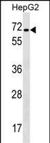
CACNB1 Antibody (C-term)
Affinity Purified Rabbit Polyclonal Antibody (Pab)
- 产品详情
- 实验流程
- 背景知识
Application
| WB, E |
|---|---|
| Primary Accession | Q02641 |
| Other Accession | Q9MZL7, NP_954855.1, NP_000714.3 |
| Reactivity | Human |
| Predicted | Bovine |
| Host | Rabbit |
| Clonality | Polyclonal |
| Isotype | Rabbit IgG |
| Calculated MW | 65714 Da |
| Antigen Region | 543-572 aa |
| Gene ID | 782 |
|---|---|
| Other Names | Voltage-dependent L-type calcium channel subunit beta-1, CAB1, Calcium channel voltage-dependent subunit beta 1, CACNB1, CACNLB1 |
| Target/Specificity | This CACNB1 antibody is generated from rabbits immunized with a KLH conjugated synthetic peptide between 543-572 amino acids from the C-terminal region of human CACNB1. |
| Dilution | WB~~1:1000 E~~Use at an assay dependent concentration. |
| Format | Purified polyclonal antibody supplied in PBS with 0.09% (W/V) sodium azide. This antibody is purified through a protein A column, followed by peptide affinity purification. |
| Storage | Maintain refrigerated at 2-8°C for up to 2 weeks. For long term storage store at -20°C in small aliquots to prevent freeze-thaw cycles. |
| Precautions | CACNB1 Antibody (C-term) is for research use only and not for use in diagnostic or therapeutic procedures. |
| Name | CACNB1 |
|---|---|
| Synonyms | CACNLB1 |
| Function | Regulatory subunit of L-type calcium channels (PubMed:1309651, PubMed:15615847, PubMed:8107964). Regulates the activity of L-type calcium channels that contain CACNA1A as pore- forming subunit (By similarity). Regulates the activity of L-type calcium channels that contain CACNA1C as pore-forming subunit and increases the presence of the channel complex at the cell membrane (PubMed:15615847). Required for functional expression L-type calcium channels that contain CACNA1D as pore-forming subunit (PubMed:1309651). Regulates the activity of L-type calcium channels that contain CACNA1B as pore-forming subunit (PubMed:8107964). |
| Cellular Location | Cell membrane, sarcolemma; Peripheral membrane protein {ECO:0000250|UniProtKB:P19517}; Cytoplasmic side {ECO:0000250|UniProtKB:P19517}. Cell membrane; Peripheral membrane protein |
| Tissue Location | Detected in heart ventricle (at protein level) (PubMed:15615847). Isoform 1 and isoform 3 are expressed in brain, heart, spleen, central nervous system and neuroblastoma cells. Isoform 2 is expressed in skeletal muscle. |
For Research Use Only. Not For Use In Diagnostic Procedures.
Provided below are standard protocols that you may find useful for product applications.
BACKGROUND
The protein encoded by this gene belongs to the calcium channel beta subunit family. It plays an important role in the calcium channel by modulating G protein inhibition, increasing peak calcium current, controlling the alpha-1 subunit membrane targeting and shifting the voltage dependence of activation and inactivation. Alternative splicing occurs at this locus and three transcript variants encoding three distinct isoforms have been identified.
REFERENCES
Jangsangthong, W., et al. Pflugers Arch. 459(3):399-411(2010)
Olsen, J.V., et al. Cell 127(3):635-648(2006)
Olsen, J.V., et al. Cell 127(3):635-648(2006)
Lim, J., et al. Cell 125(4):801-814(2006)
Foell, J.D., et al. Physiol. Genomics 17(2):183-200(2004)
终于等到您。ABCEPTA(百远生物)抗体产品。
点击下方“我要评价 ”按钮提交您的反馈信息,您的反馈和评价是我们最宝贵的财富之一,
我们将在1-3个工作日内处理您的反馈信息。
如有疑问,联系:0512-88856768 tech-china@abcepta.com.


