


 癌症的基本特征包括细胞增殖、血管生成、迁移、凋亡逃避机制和细胞永生等。找到癌症发生过程中这些通路的关键标记物和对应的抗体用于检测至关重要。
 为您推荐一个泛素化位点预测神器——泛素化分析工具,可以为您的蛋白的泛素化位点作出预测和评分。
 细胞自噬受体图形绘图工具为你的蛋白的细胞受体结合位点作出预测和评分,识别结合到自噬通路中的蛋白是非常重要的,便于让我们理解自噬在正常生理、病理过程中的作用,如发育、细胞分化、神经退化性疾病、压力条件下、感染和癌症。
ALDH1A1 Antibody
Affinity Purified Rabbit Polyclonal Antibody (Pab)
- 产品详情
- 文献引用 : 1
- 实验流程
- 背景知识
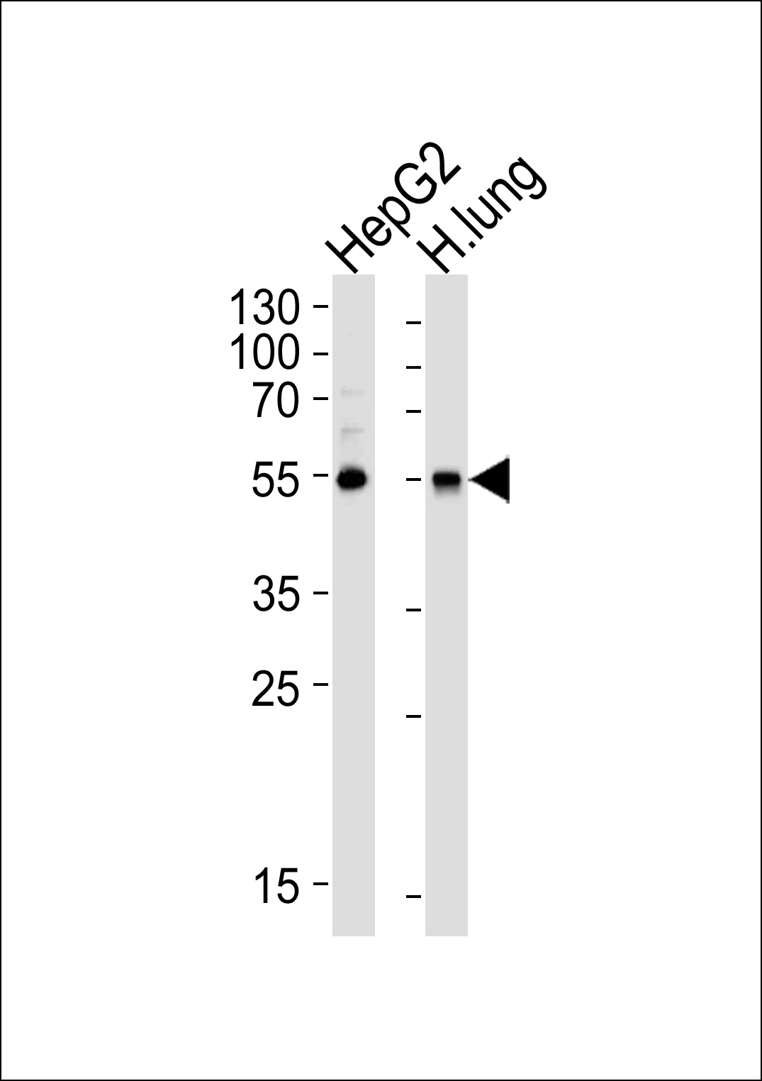
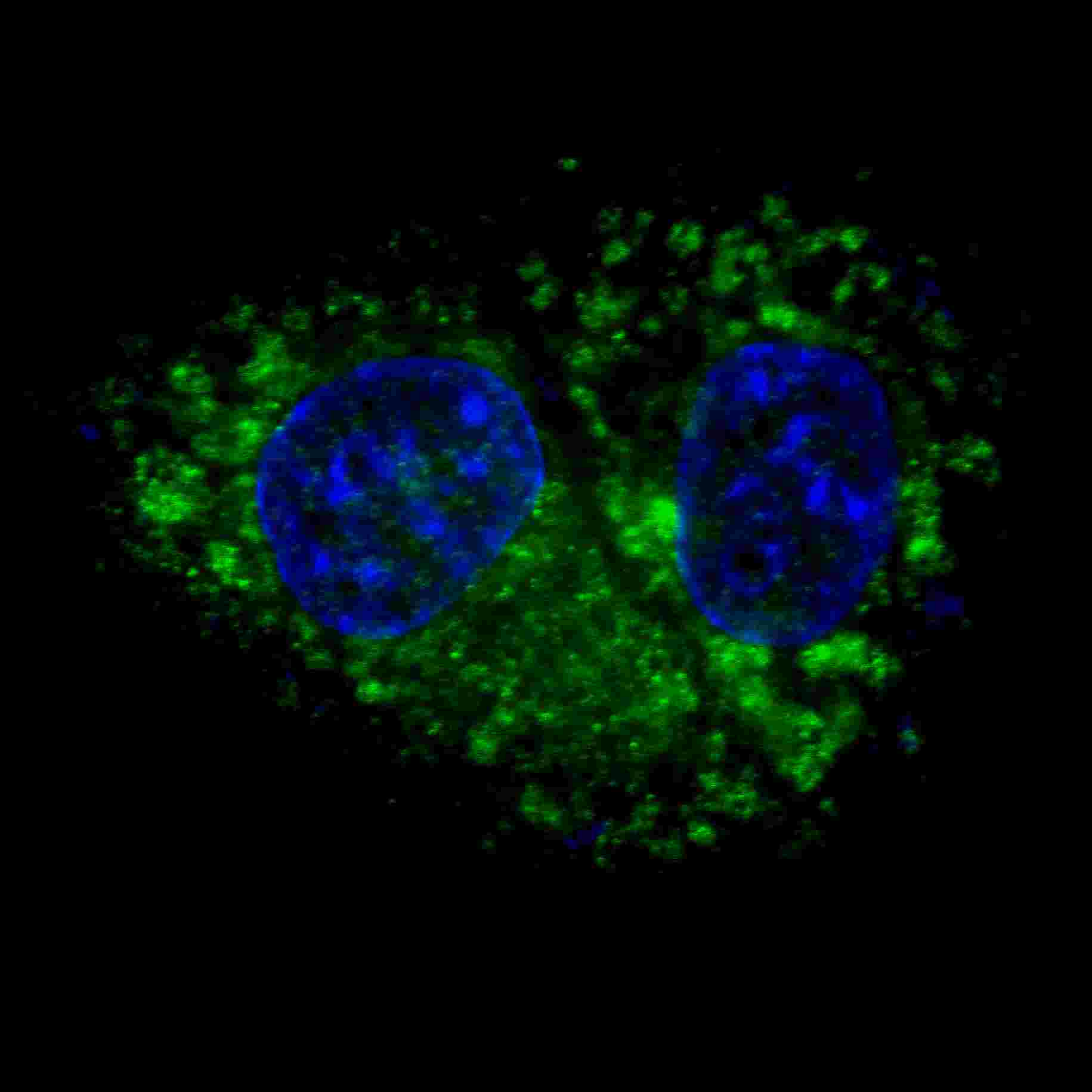
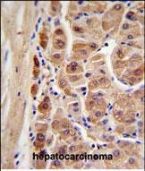
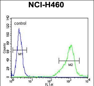
Application
| IF, IHC-P, FC, WB, E |
|---|---|
| Primary Accession | P00352 |
| Reactivity | Human |
| Host | Rabbit |
| Clonality | Polyclonal |
| Isotype | Rabbit IgG |
| Calculated MW | 54862 Da |
| Gene ID | 216 |
|---|---|
| Other Names | Retinal dehydrogenase 1, RALDH 1, RalDH1, ALDH-E1, ALHDII, Aldehyde dehydrogenase family 1 member A1, Aldehyde dehydrogenase, cytosolic, ALDH1A1, ALDC, ALDH1, PUMB1 |
| Target/Specificity | This ALDH1A1 antibody is generated from rabbits immunized with human ALDH1A1 recombinant protein. |
| Dilution | IF~~1:100 IHC-P~~1:100~500 FC~~1:10~50 WB~~1:1000 E~~Use at an assay dependent concentration. |
| Format | Purified polyclonal antibody supplied in PBS with 0.09% (W/V) sodium azide. This antibody is purified through a protein A column, followed by peptide affinity purification. |
| Storage | Maintain refrigerated at 2-8°C for up to 2 weeks. For long term storage store at -20°C in small aliquots to prevent freeze-thaw cycles. |
| Precautions | ALDH1A1 Antibody is for research use only and not for use in diagnostic or therapeutic procedures. |
| Name | ALDH1A1 (HGNC:402) |
|---|---|
| Function | Cytosolic dehydrogenase that catalyzes the irreversible oxidation of a wide range of aldehydes to their corresponding carboxylic acid (PubMed:12941160, PubMed:15623782, PubMed:17175089, PubMed:19296407, PubMed:25450233, PubMed:26373694). Functions downstream of retinol dehydrogenases and catalyzes the oxidation of retinaldehyde into retinoic acid, the second step in the oxidation of retinol/vitamin A into retinoic acid (By similarity). This pathway is crucial to control the levels of retinol and retinoic acid, two important molecules which excess can be teratogenic and cytotoxic (By similarity). Also oxidizes aldehydes resulting from lipid peroxidation like (E)-4-hydroxynon-2-enal/HNE, malonaldehyde and hexanal that form protein adducts and are highly cytotoxic. By participating for instance to the clearance of (E)-4-hydroxynon-2-enal/HNE in the lens epithelium prevents the formation of HNE-protein adducts and lens opacification (PubMed:12941160, PubMed:15623782, PubMed:19296407). Also functions downstream of fructosamine-3-kinase in the fructosamine degradation pathway by catalyzing the oxidation of 3-deoxyglucosone, the carbohydrate product of fructosamine 3-phosphate decomposition, which is itself a potent glycating agent that may react with lysine and arginine side-chains of proteins (PubMed:17175089). Also has an aminobutyraldehyde dehydrogenase activity and is probably part of an alternative pathway for the biosynthesis of GABA/4-aminobutanoate in midbrain, thereby playing a role in GABAergic synaptic transmission (By similarity). |
| Cellular Location | Cytoplasm, cytosol. Cell projection, axon {ECO:0000250|UniProtKB:P24549} |
| Tissue Location | Expressed by erythrocytes (at protein level). |
For Research Use Only. Not For Use In Diagnostic Procedures.

Provided below are standard protocols that you may find useful for product applications.
BACKGROUND
ALDH1A1 belongs to the aldehyde dehydrogenases family of proteins. Aldehyde dehydrogenase is the second enzyme of the major oxidative pathway of alcohol metabolism. Two major liver isoforms of this enzyme, cytosolic and mitochondrial, can be distinguished by their electrophoretic mobilities, kinetic properties, and subcellular localizations. Most Caucasians have two major isozymes, while approximately 50% of Orientals have only the cytosolic isozyme, missing the mitochondrial isozyme. A remarkably higher frequency of acute alcohol intoxication among Orientals than among Caucasians could be related to the absence of the mitochondrial isozyme.
REFERENCES
References for protein:
1.Moore,S., J Stud Alcohol Drugs 68 (2), 192-196 (2007)
2.Collard,F., Biochimie 89 (3), 369-373 (2007)
References for HepG2 cell line:
1. Knowles BB, et al. (1980). Human hepatocellular carcinoma cell lines secrete the major plasma proteins and hepatitis B surface antigen. Science 209: 497-499.[ PubMed: 6248960].
2. Darlington GJ, et al. (1987). Growth and hepatospecific gene expression of human hepatoma cells in a defined medium. In Vitro Cell. Dev. Biol. 23: 349-354.[PubMed: 3034851].
3. Ihrke, G; Neufeld, EB; Meads, T; Shanks, MR; Cassio, D; Laurent, M; Schroer, TA; Pagano, RE et al. (1993). "WIF-B cells: an in vitro model for studies of hepatocyte polarity". Journal of Cell Biology 123 (6): 1761–1775. [PubMed:7506266].
4. Mersch-Sundermann, V.; Knasmüller, S.; Wu, X. J.; Darroudi, F.; Kassie, F. (2004). "Use of a human-derived liver cell line for the detection of cytoprotective, antigenotoxic and cogenotoxic agents". Toxicology 198 (1–3): 329–340. [PubMed:15138059].
终于等到您。ABCEPTA(百远生物)抗体产品。
点击下方“我要评价 ”按钮提交您的反馈信息,您的反馈和评价是我们最宝贵的财富之一,
我们将在1-3个工作日内处理您的反馈信息。
如有疑问,联系:0512-88856768 tech-china@abcepta.com.


