


 癌症的基本特征包括细胞增殖、血管生成、迁移、凋亡逃避机制和细胞永生等。找到癌症发生过程中这些通路的关键标记物和对应的抗体用于检测至关重要。
 为您推荐一个泛素化位点预测神器——泛素化分析工具,可以为您的蛋白的泛素化位点作出预测和评分。
 细胞自噬受体图形绘图工具为你的蛋白的细胞受体结合位点作出预测和评分,识别结合到自噬通路中的蛋白是非常重要的,便于让我们理解自噬在正常生理、病理过程中的作用,如发育、细胞分化、神经退化性疾病、压力条件下、感染和癌症。
WIPI2 Antibody (N-term)
Affinity Purified Rabbit Polyclonal Antibody (Pab)
- 产品详情
- 文献引用 : 1
- 实验流程
- 背景知识
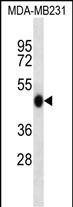
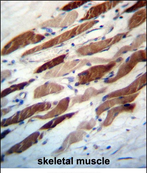
Application
| WB, IHC-P, E |
|---|---|
| Primary Accession | Q9Y4P8 |
| Other Accession | NP_001028690.1, NP_057087.2, NP_001028691.1 |
| Reactivity | Human |
| Host | Rabbit |
| Clonality | Polyclonal |
| Isotype | Rabbit IgG |
| Calculated MW | 49408 Da |
| Antigen Region | 4-32 aa |
| Gene ID | 26100 |
|---|---|
| Other Names | WD repeat domain phosphoinositide-interacting protein 2, WIPI-2, WIPI49-like protein 2, WIPI2 |
| Target/Specificity | This WIPI2 antibody is generated from rabbits immunized with a KLH conjugated synthetic peptide between 4-32 amino acids from the N-terminal region of human WIPI2. |
| Dilution | WB~~1:1000 IHC-P~~1:100~500 E~~Use at an assay dependent concentration. |
| Format | Purified polyclonal antibody supplied in PBS with 0.09% (W/V) sodium azide. This antibody is purified through a protein A column, followed by peptide affinity purification. |
| Storage | Maintain refrigerated at 2-8°C for up to 2 weeks. For long term storage store at -20°C in small aliquots to prevent freeze-thaw cycles. |
| Precautions | WIPI2 Antibody (N-term) is for research use only and not for use in diagnostic or therapeutic procedures. |
| Name | WIPI2 (HGNC:32225) |
|---|---|
| Function | Component of the autophagy machinery that controls the major intracellular degradation process by which cytoplasmic materials are packaged into autophagosomes and delivered to lysosomes for degradation (PubMed:20505359, PubMed:28561066). Involved in an early step of the formation of preautophagosomal structures (PubMed:20505359, PubMed:28561066). Binds and is activated by phosphatidylinositol 3- phosphate (PtdIns3P) forming on membranes of the endoplasmic reticulum upon activation of the upstream ULK1 and PI3 kinases (PubMed:28561066). Mediates ER-isolation membranes contacts by interacting with the ULK1:RB1CC1 complex and PtdIns3P (PubMed:28890335). Once activated, WIPI2 recruits at phagophore assembly sites the ATG12-ATG5-ATG16L1 complex that directly controls the elongation of the nascent autophagosomal membrane (PubMed:20505359, PubMed:28561066). |
| Cellular Location | Preautophagosomal structure membrane; Peripheral membrane protein; Cytoplasmic side. Note=Localizes to omegasomes membranes which are endoplasmic reticulum connected structures at the origin of preautophagosomal structures. Enriched at preautophagosomal structure membranes in response to PtdIns3P. |
| Tissue Location | Ubiquitously expressed (at protein level). Highly expressed in heart, skeletal muscle and pancreas. Expression is down- regulated in pancreatic and in kidney tumors |
For Research Use Only. Not For Use In Diagnostic Procedures.

Provided below are standard protocols that you may find useful for product applications.
BACKGROUND
WD40 repeat proteins are key components of many essential biologic functions. They regulate the assembly of multiprotein complexes by presenting a beta-propeller platform for simultaneous and reversible protein-protein interactions. Members of the WIPI subfamily of WD40 repeat proteins, such as WIPI2, have a 7-bladed propeller structure and contain a conserved motif for interaction with phospholipids (Proikas-Cezanne et al., 2004 [PubMed 15602573]).
REFERENCES
Sugiyama, N., et al. Mol. Cell Proteomics 6(6):1103-1109(2007)
Proikas-Cezanne, T., et al. Oncogene 23(58):9314-9325(2004)
Simpson, J.C., et al. EMBO Rep. 1(3):287-292(2000)
终于等到您。ABCEPTA(百远生物)抗体产品。
点击下方“我要评价 ”按钮提交您的反馈信息,您的反馈和评价是我们最宝贵的财富之一,
我们将在1-3个工作日内处理您的反馈信息。
如有疑问,联系:0512-88856768 tech-china@abcepta.com.


