


 癌症的基本特征包括细胞增殖、血管生成、迁移、凋亡逃避机制和细胞永生等。找到癌症发生过程中这些通路的关键标记物和对应的抗体用于检测至关重要。
 为您推荐一个泛素化位点预测神器——泛素化分析工具,可以为您的蛋白的泛素化位点作出预测和评分。
 细胞自噬受体图形绘图工具为你的蛋白的细胞受体结合位点作出预测和评分,识别结合到自噬通路中的蛋白是非常重要的,便于让我们理解自噬在正常生理、病理过程中的作用,如发育、细胞分化、神经退化性疾病、压力条件下、感染和癌症。
Mouse Monoclonal Antibody to CTNNA1
Purified Mouse Monoclonal Antibody
- 产品详情
- 实验流程
Application
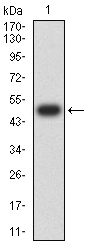
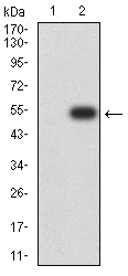
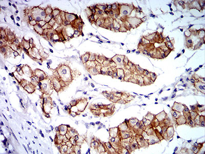
| WB, IHC, E |
|---|---|
| Primary Accession | P35221 |
| Reactivity | Human |
| Host | Mouse |
| Clonality | Monoclonal |
| Clone Names | 8B6C1 |
| Isotype | Mouse IgG1 |
| Calculated MW | 100071 Da |
| Description | CTNNA1 (Catenin (Cadherin-Associated Protein), Alpha 1, 102kDa) is a Protein Coding gene. Diseases associated with CTNNA1 include diffuse gastric cancer and acquired thrombocytopenia. Among its related pathways are Signaling by GPCR and Developmental Biology. GO annotations related to this gene include poly(A) RNA binding and actin filament binding. An important paralog of this gene is CTNNA3.; |
| Immunogen | Purified recombinant fragment of human CTNNA1 (AA: 371-574) expressed in E. Coli. |
| Formulation | Purified antibody in PBS with 0.05% sodium azide |
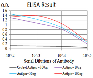
| Application Note | ELISA: 1/10000; WB: 1/500 - 1/2000; IHC: 1/200 - 1/1000; |
| Gene ID | 1495 |
|---|---|
| Other Names | CAP102 |
| Dilution | WB~~1:1000 IHC~~1:100~500 E~~N/A |
| Storage | Maintain refrigerated at 2-8°C for up to 6 months. For long term storage store at -20°C in small aliquots to prevent freeze-thaw cycles. |
| Precautions | Mouse Monoclonal Antibody to CTNNA1 is for research use only and not for use in diagnostic or therapeutic procedures. |
| Name | CTNNA1 (HGNC:2509) |
|---|---|
| Function | Associates with the cytoplasmic domain of a variety of cadherins. The association of catenins to cadherins produces a complex which is linked to the actin filament network, and which seems to be of primary importance for cadherins cell-adhesion properties. Can associate with both E- and N-cadherins. Originally believed to be a stable component of E-cadherin/catenin adhesion complexes and to mediate the linkage of cadherins to the actin cytoskeleton at adherens junctions. In contrast, cortical actin was found to be much more dynamic than E-cadherin/catenin complexes and CTNNA1 was shown not to bind to F-actin when assembled in the complex suggesting a different linkage between actin and adherens junctions components. The homodimeric form may regulate actin filament assembly and inhibit actin branching by competing with the Arp2/3 complex for binding to actin filaments. Involved in the regulation of WWTR1/TAZ, YAP1 and TGFB1- dependent SMAD2 and SMAD3 nuclear accumulation (By similarity). May play a crucial role in cell differentiation. |
| Cellular Location | Cytoplasm, cytoskeleton {ECO:0000250|UniProtKB:P26231}. Cell junction, adherens junction. Cell membrane {ECO:0000250|UniProtKB:P26231}; Peripheral membrane protein; Cytoplasmic side {ECO:0000250|UniProtKB:P26231}. Cell junction Cytoplasm {ECO:0000250|UniProtKB:Q9PVF8}. Nucleus. Note=Found at cell-cell boundaries and probably at cell-matrix boundaries. {ECO:0000250|UniProtKB:P26231} |
| Tissue Location | [Isoform 1]: Ubiquitously expressed in normal tissues. |
Research Areas
For Research Use Only. Not For Use In Diagnostic Procedures.
Application Protocols
Provided below are standard protocols that you may find useful for product applications.
REFERENCES
1.Cell Cycle. 2014;13(15):2334-9. ; 2.J Pathol. 2013 Mar;229(4):621-9. ;
终于等到您。ABCEPTA(百远生物)抗体产品。
点击下方“我要评价 ”按钮提交您的反馈信息,您的反馈和评价是我们最宝贵的财富之一,
我们将在1-3个工作日内处理您的反馈信息。
如有疑问,联系:0512-88856768 tech-china@abcepta.com.
¥ 1,500.00
Cat# AO2373a


